Download
1 / 83
960 likes | 1.71k Views
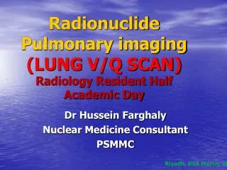
Radionuclide Pulmonary imaging (LUNG V/Q SCAN) Radiology Resident Half Academic Day. Dr Hussein Farghaly Nuclear Medicine Consultant PSMMC. Riyadh, KSA March, 2011. Clinical Application:

E N D
Radionuclide Pulmonary imaging(LUNG V/Q SCAN)Radiology Resident Half Academic Day Dr Hussein Farghaly Nuclear Medicine Consultant PSMMC Riyadh, KSA March, 2011
Clinical Application: • A. The most common clinical indication for lung scintigraphy is to determine the likelihood of pulmonary embolism (PE). • B. Less common clinical indications are: 1. Document the degree of resolution of pulmonary embolism 2. Quantify differential pulmonary function before pulmonary surgery for lung cancer 3. Evaluate lung transplants 4. Evaluate congenital heart or lung disease such as cardiac shunts, pulmonary arterial stenosis, and arteriovenous fistulae and their treatment 5. Confirm the presence of bronchopleural fistula 6. Evaluate chronic pulmonary parenchymal disorders such as cystic fibrosis 7. Evaluate the cause of pulmonary hypertension
PROCEDURE/SPECIFICATIONS OF THE EXAMINATION • Nuclear Medicine study request: -Women of childbearing age -Clinical pre test probability of PE (Wells score) -D-Dimer test -History of prior DVT or PE or anticoagulant therapy -Review of prior lung scintigraphy -Relevant chest radiographic findings • Patient Preparation and Precautions -A standard chest X-ray CT scan can substitute - Pregnant or breastfeeding women: benefit versus risk. - Discontinue breastfeeding for 24 hours - Children < 3 years of age: Limit close contact for 24 hours.
Perfusion scan: Agent: • Tc-99m macro-aggregated albumin (Tc-99m MAA) • Particle size: 10-90 microns (90% of particles) • No particles should be larger than 150 microns • Obstructing ~ 0.1% of precapillary arterioles • Particles clearance: Enzymatic hydrolysis and phagocytized by reticuloendothelial cells (biologic half-life of 6-8 hours). • Critical organ: Lungs, 1 rad/5 mCi Total RDE : ~ 0.8 mSv
Perfusion scan: Agentcont.: Number of particles: 500,000 particles/5mCi, Minimum of 70,000 Reduced number of particles: 100,000 in Pulmonary arterial hypertension (> 50 mmHg), right to left shunts, status post pneumonectomy, poor respiratory function, cardiopulmonary instability. 10,000 to 50,000 particles in Neonates: • Neonate have about 10% of adult pulmonary capillaries • Number of capillaries increases to half of the adult value by age 3 years, and reaches an adult level by age 8 to 12 years. • Dose and adminstraion: Adult 40–150 MBq (1–4 mCi). Pediatric 1.11 MBq/kg (0.03 mCi/kg) with a minimum of 14.8 MBq/kg (0.4 mCi) - Tc-99m MAA is injected IV slowly during 3–5 respiratory cycles with the patient in the supine position.. -
Perfusion scan: Imaging • Imaging is preferably performed in the upright position to increase chest cavity size and to minimize diaphragmatic motion. • Large field of view camera, parallel hole and high-resolution collimator. Planar images should be obtained in multiple projections including anterior, posterior, both posterior oblique, both anterior oblique and both lateral projections • SPECT can be used to obtain a three-dimensional evaluation of the perfusion, and is recommended by some investigators. • Images of the brain may be obtained to distinguish right-to-left shunting from systemic distribution of radiopharmaceutical components too small to be trapped by capillaries. • Tc-99m MAA injection into the veins of the feet: May help to reveal a DVT as "hot spots" Venous obstruction or collateral flow can be seen
Ventilation scan: Radiopharmaceuticals RADIOACTIVE GASES: - Xenon-133 (5 days, 81KeV ) - Krypton-81m (13 sec, 190 KeV, RU-81/Kr-81m generator) RADIOAEROSOL: -Technegas “pseudo-gas (0.005–0.2 μm) -Tc-99m DTPA aerosol (0.1–0.5 μm)
Ventilation scan:Xenon-133 • Gamma energy: 81 keV • Physical half-life: 5.2 days • Dose inhaled: 15-25 mCi through Xenon delivery device. • Critical organs: Trachea and airways of ~ 250 mrad • Absorbed dose by the body: Less than 15% • Total radiation dose equivalent: 1.2 -2.0 mSv • Xe-133 ventilation scan performed prior to Q scan • Uncommon practice: Xe-133 exam after the perfusion study; low dose of Tc-99m MAA first (1-2 mCi) and then high dose of Xe-133 (20-30 mCi)
Ventilation scan:Xenon-133 • Gamma camera placed behind the patient's back: Posterior view. • Xe-133 is inhaled through a mouthpiece of the Xenon delivery system. Ventilation scan in 3 phases: • Single breath: At maximum inspiration take 10-15 sec image. COPD detected in ~ 66% • Equilibrium: Normal tidal respirations for 3-5 minutes, rebreathing a mixture of Xenon-133 and oxygen take two sequential 90 sec images. Least sensitive for COPD diagnosis. • Washout: Patient breaths room air or oxygen while exhaling the xenon into a charcoal trap take three sequential 45 sec images. Biological half life is 30 sec. • Normal Xenon-133 clearance: 2-3 minutes. Clearance > 3 minutes: Air trapping. More sensitive (90%) than single breath phase in diagnosing COPD. • Xenon more sensitive than aerosolized Tc-DTPA for detecting COPD.
Ventilation scan: Krypton-81m • Rubidium krypton-81m generator. • Isomeric transition like Tc-99m • Gamma emission: 191 keV (65%) • Physical half-life: 13 seconds • Advantages: • Higher energy than Tc-99m • Very short physical half-life: V imaging immediately after each Q image • Radiation dose to the lungs is lower than with other agents (about 15 mrad per view). • Disadvantages: • Rubidium 81m: Physical half-life ~ 4.5 hours, can be used only for 1 day • Limited assessment of COPD because tracer decays before an equilibrium distribution can be attained
Ventilation scan: Tc-99m DTPA • Submicronic particles (aerosols) inhaled through an aerosol delivery system. • Particles size: 0.5 to 0.8 microns in diameter, reaching alveoli • Dose: 30 mCi (3-5 minutes rebreathing within closed system, oxygen at 8-10 liters/min). • System delivers about 0.5-0.8 mCi of tracer to the lungs 100,000 counts images in about 2-5 minutes. • Radiation exposure to the lungs: ~ 100 mrads. • Exposure to personnel: Less compared with Xenon study. • Half-time clearance: 1-1.5 hours in normal patients; more rapid clearance in patients with PE, lung injury (ARDS), pulmonary fibrosis, and in smokers (20 min.)
Tc-99m DTPA vs. Xe-133 Advantages of Tc-99m DTPA : • No collection devices for exhaled gas • Multiple projections • Can perform before or after perfusion scan • Feasible for ICU patients on ventilator Disadvantage of Tc-99m DTPA : • Air trapping cannot be assessed. • Xenon more sensitive than Tc-DTPA for detecting COPD.
Ventilation scan: Tc-99m technegas • Tc-04 is vaporized in a microfurnace: Ultrafine labelled carbon particles. • Particle size: 0.05 and 0.15 microns • Good peripheral deposition: even in COPD. • Longer pulmonary retention no effective clearance half time is 6hrs • The material is produced by heating 5mCi of Tc- pertechnetate to very high temperatures (2500 degrees Celsius) in the presence of 100% argon gas produced a Tc-carbon particle that is so small it acts like a gas.
Image Sequence: • A. A chest radiograph should be obtained and reviewed before lung scintigraphy. • B. Ventilation scintigraphy using 133Xe is usually performed before perfusion scintigraphy. Alternately, perfusion scintigraphy can be performed first and ventilation scintigraphy omitted if not needed. • C. Because of the higher energy of the gamma emissions and the short half-life of 81mKr, images obtained with this gas can be alternated with those obtained with 99mTc MAA. • D. When 99mTc labeled aerosol imaging is performed before 99mTc MAA perfusion imaging, smaller amounts (20-40 MBq) [0.5-1.0 mCi]) of 99mTc labeled aerosol should be administered to the lungs.
ACUTE PULMONARY EMBOLISM CLINICAL PRESENTATION: (Non-specific) • Haemoptysis, Dyspnea and Pleuritic Chest pain (Virchows triad) • Back or Abdominal pain, cough, SOB, Low-grade fever,---------- • May be asympotmatic
Evaluation • ABG – Respiratory alkalosis, hypoxia • ECG – Sinus tachycardia & S1Q3T3 • D-Dimer • CXR • Spiral CT with contrast • V/Q Scan • Angiogram
Question 1 Pulmonary angiography as “gold standard” Sensitivity for PE is: • 97% • 93% • 87%
Question 2 Accuracy of V/Q scan in PIOPED – incorrect answer? • 98% sensitivity • 10% specificity • High-probability V/Q scans as PE criteria: Failed to detect PE in 59% of patients • 70% specificity
Question 3 Accuracy of multiple slice CTA – incorrect answer? • Variable sensitivities from 53% to 87% in different studies • Reader’s experience is important • Specificity > 90% • Sensitivity is higher than specificity
Question 4 Diagnostic accuracy of CTA – incorrect answer? • Dependent on clinical probability for PE • CTA has high NPV similar to that at V/Q scan • Independent from clinical probability for PE
Diagnostic Pathways in Acute Pulmonary EmbolismRecommendations of The PIOPED II Investigators
Pre Imaging Objective clinical probability • Three clinical scoring system have been tested prospectively and validated in large scale clinical trials: Wells’ score (Ann Intern Med 1998) Geneva Score (Arch Intern Med 2001, Ann Intern Med 2006) Pisa Score (Ann RespirCrit Care Med 1999, Ann j Med 2003)
The diagnostic yield of D-Dimer is lower in cancer patient, the elderly, inpatient, recent trauma or surgery and during pregnancy
CHEST X- Ray • Initial CXR usually normal. • May progress to show atelectasis, plueral effusion and elevated hemidiaphram. • Hampton’s hump and Westermark signs are classic findings but are not usually present.
Hampton’s Hump – consists of a pleura based shallow wedge-shaped consolidation in the lung periphery with the base against the pleural surface. • Westermark sign – Dilatation of pulmonary vessels proximal to embolism along with collapse of distal vessels, often with a sharp cut off.
Lung V/Q scan • Should lung scan be omitted for pulmonary embolism diagnosis in the age of multislice spiral CT? A) YES B) NO NO, Lung scan has a role in PE diagnosis When there are: Contraindications to CT Scan: Allergy to iodinated contrast agent Renal failure Pregnancy? High diagnostic yield and avoidance of unnecessary radiation exposure. Pregnancy Young patient with normal X-ray.
Interpretation Criteria of V/Q scan - Prospective Investigation of Pulmonary Embolism Diagnosis (PIOPED), 1990 • Revised PIOPED, 1995 • PISA-PED, 1996: Perfusion scan only • PIOPED II , 2006 • Modified PIOPED II : perfusion and CXR
PIOPED • 933/1,493 patients analyzed • 755 of these patients with pulmonary angiography within 12– 24 h of V/Q scan • Posterior xenon-133 ventilation scan, followed by an 8-view Tc-99m MAA perfusion lung scan • One-year follow-up: New PE, major bleeding complications, or death 1Value of the ventilation/perfusion scan in acute pulmonary embolism. Results of the Prospective Investigation of Pulmonary Embolism Diagnosis (PIOPED). The PIOPED Investigators. JAMA 1990; 263:2753-9
V/Q scan accuracy: PIOPED • Based on PA: 98% sensitivity and 10% specificity for V/Q scan • High-probability V/Q scans (V/Q mismatch) as criteria for PE: Failed to detect PE in 59% of patients, based on PA.
Likelihood of PE: PIOPED Predictive values > 90%: Only 22% of patients. Combined V/Q scan and clinical probability: Highest diagnostic accuracy. High clinical probability & high-probability V/Q scan: 95% likelihood of PE. Low clinical probability & low-probability V/Q scan: 4% likelihood of PE.
PISA-PED, 1996: Perfusion scan only • 890 patients with Q scan, compared with PA • 413/670 (62%) patients with abnormal Q scans had PA; no PA if normal/near normal Q scan • 92% sensitivity and 87% specificity • Positive Q scan and high clinical suspicion: PPV >90% • Negative Q scan and low clinical suspicion: NPV of 97%.
Pisa Ped perfusion scan categories and interpretation criteria Miniati M, et al: Value of perfusion lung scan in the diagnosis of pulmonary embolism: Results of the Prospective Investigative Study of Acute Pulmonary Embolism Diagnosis (PISA-PED). Am J Respir Crit Care Med 1996;154:1387–1393.
PISA-PED: Conclusion • Q scanning alone: Much closer to angiography than V/Q scanning • Q scanning rather than V/Q scanning: Imaging technique of first choice for diagnosis of PE
PIOPED II: V/Q scan results • PE present or PE absent: 74% (PISA-PED: 75%) • Sensitivity for PE present: 77% (CTA: 83%) • Specificity of PE absent: 98% (CTA: 98%) Conclusions: V/Q scan provides definitive diagnosis in a majority of patients (74%) Sostman HD, et al. Acute pulmonary embolism: sensitivity and specificity of ventilation perfusion scintigraphy in PIOPED II study. Radiology 2008; 246: 941-946
Stripe Sign: A thin line (stripe) of activity between a Q defect and adjacent pleural surface: sometime in emphysema. Only 6% prevalence of PE.Triple match: Matching Q and V defect, and CXR abnormality, regardless of size: Atelectasis, consolidation. Prevalence of PE: 26% (upper - 11%; middle - 12%; lower - 33%)1