Download
1 / 71
710 likes | 1.42k Views
ADULT RESPIRATORY DISTRESS. DAN MUSE, MD. Conditions that result in a compromise in oxygenation and ventilation . Can result in an increase or decrease in respiratory drive. RESPIRATORY DISTRESS. OXYGENATION: Providing oxygen to the cells in the body.

E N D
ADULT RESPIRATORY DISTRESS DAN MUSE, MD
Conditions that result in a compromise in oxygenation and ventilation. • Can result in an increase or decrease in respiratory drive. RESPIRATORY DISTRESS
OXYGENATION: Providing oxygen to the cells in the body. • VENTILATION: The exchange of CO2 for oxygen. OXYGENATION - VENTILATION
Results from fluid building up into the lungs through hydrostatic pressure and changes in osmotic pressure. • The pressure of the blood and plasma in the vessels traversing the lungs increases to the point where by the plasma fluid leaches out into the lungs. CONGESTIVE HEART FAILURE
Oncotic pressure “pulls in”. • Large molecules such as albumin and other proteins need fluid and draw it in from the soft tissue. • A change in these pressures i.e. ”low albumin” can cause fluids to leach back out. Oncotic pressure
Hydrostatic pressure “pushes out” • Fluids in the vessel leach out in order to equilibrate with the lower outside pressure. HYDROSTATIC PRESSURE
CAUSES MAY INCLUDE……….. • Pump failure. The heart no longer has the strength to pump effectively. Cardiomegally and low ejection fraction. • Valvular heart disease resulting in backwashing • Vascular resistance causing resistance to outflow CONGESTIVE HEART FAILURE
SYMPTOMS… • Shortness Of Breath • Pnd And Orthopnea • Peripherial Edema CONGESTIVE HEART FAILURE
CLINICAL SIGNS…… • Increased Respiratory Rate • Bp Can Be Low, Normal Or High • Rales, Diminished Breath Sounds, Wheezes • JVD, Peripheral Edema CONGESTIVE HEART FAILURE
TREATMENT…… • OXYGEN…….Increases oxygen carried in the cells • LASIX……Removes fluid from the body. This in turn reduces the hydrostatic pressure in the vessels and makes the amount of blood more manageable. • NITRITES…….Dilates the vessels so that they can accommodate the fluid. • CPAP…… CONGESTIVE HEART FAILURE
RADIOGRAPH OF CHFENLARGED HEARTFLUID (THE WHITE) BUILD UP IN THE LUNGS
Really Bad CHF • Most Often Associated With Cardiac Ischemia • Typically Has A Sudden Onset. • Findings Early On Include Hypertension, And Rales. PULMONARY EDEMA
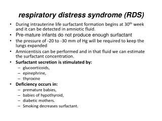
ALSO KNOWN AS ARDS…Adult Respiratory Distress Syndrome • Distinguished from pulmonary edema by underlying cause. • Typically the heart size is normal. • Is a symptom of another problem. NONCARDIOGENIC PULMONARY EDEMA
CAUSES….. • Head Injury • Infection • Drugs • Severe Trauma • Smoke Inhalation And Burns • Inflamatory Processes Such As Pancreatitis NONCARDIOGENIC PULMONARY EDEMA
SYMPTOMS….. • Similar to CHF and pulmonary edema. • SOB and often sudden. NONCARDIOGENIC PULMONARY EDEMA
CLINICAL FINDINGS….. • SOB with rales. • Blood pressure variable NONCARDIOGENIC PULMONARY EDEMA
TREATMENT…… • Supportive. Need to treat underlying cause • Diuretics typically do not work • You will give the diuretic because you can’t distinguish noncardiogenic from cardiogenic in the field. NONCARDIOGENIC PULMONARY EDEMA
NONCARDIOGENIC PULMONARY EDEMA CAN NOT DISTINGUISH FROM CARDIOGENIC PULMONARY EDEMA
Caused By A Clot Lodging In A Pulmonary Vessel PULMONARY EMBOLUS
CAUSES………. • Typically associated with recent surgery, trauma or travel. • Higher rate in women who smoke and take bcp’s. • May be first time presentation for someone with cancer or blood disorder.
SYMPTOMS • Symptoms will vary depending on the size and location of the clot. • May have pleuritic chest pain caused by lack of blood flow to the peripheral lung near the pleura. • Shortness of breath • Swollen leg if DVT is a source PULMONARY EMBOLUS
CLINICAL SIGNS…… • Increased respiratory rate. • Lungs may be clear, diminished or even wheezes • Heart rate may be elevated. Most common finding is tachycardia • Leg swelling PULMONARY EMBOLUS
EKG….. • Can result in finding of right sided heart strain because of the outflow blockage from the right ventricle • Most common finding is tachycardia • May have S1, Q3,T3 • Can cause incomplete or complete right bundle. PULMONARY EMBOLUS
TREATMENT…… • Oxygen • Treatment is predicated on clinical findings. • Can vary from transporting to CPR PULMONARY EMBOLUS
POTENTIAL FINDINGS ON EKG OF A PE • S1Q3T3 • RIGHT INCOMPLETE OR COMPLETE BUNDLE
THROMBUS IN MAIN PULMONARY ARTERY THE MORE PROXIMAL TO THE HEART THE MORE LETHAL
WHEN AIR OCCUPIES THE PLEURAL SPACE CAUSING COMPRESSION OF THE LUNG PNEUMOTHORAXPTX
CAUSES……… • Seen in thoracic trauma. • May be caused by penetration into the thorax or from sudden changes in pressure on the lungs • Spontaneous causes • May result from space occupying lesions in the chest PNEUMOTHORAXPTX
SYMPTOMS…… • Varying degree of shortness of breath. • Splinting and/or pain on inspiration • Tachycardia • Chest wall pain if traumatic PNEUMOTHORAX
CLINICAL SIGNS…… • Normal to elevated respiratory rate • Normal to rapid heart rate • Normal to diminished breath sounds. • Chest wall tenderness and/or crepitus • Tracheal deviation PNEUMOTHORAX
PNEUMOTHORAXCT SCAN CAUSED BY LUNG PUNCTURE FROM CENTRAL LINE
CLINICAL SIGNS • Elevated respiratory and heart rate • Change in blood pressure…usually down • Very dyspneic • Tracheal deviation may be present • Crepitus may be present. • Diminished to loss of breath sounds TENSION PNEUMOTHORAX
TENSION PNEUMOTHORAX Note The Pressure Caused By The Pneumothorax Compressing Everything To The Left.
TREATMENT……. • Supportive measures if no respiratory compormise • If clinically the patient has a tension ptx, this is a life threatening event and the patient needs immediate intervention…NEEDLE THORACENTESIS PNEUMOTHORAX
CHRONIC OBSTRUCTIVE PULMONARY DISEASE • Made up of emphysema, chronic bronchitis and asthma. • Results from inflammation of the bronchioles which causes narrowing. • This in turn causes difficulty for air to pass in and out. COPD
CAUSES……. • Smoking • Smoking • Asbestosis • 2nd Hand Smoke • Restrictive Lungs • Reactive Airway Disease • Allergic And Exercise Induced COPD
SYMPTOMS……. • Increasing difficulty breathing • Oftentimes associated with upper respiratory infections • Asthmatics may have a cough equivalent COPD