Cerebral Cortex
Cerebral Cortex. I. Organization II. Afferent/Efferent Connections III. Brodmann’s Functional areas IV. Neurological Consequences of Cortical Focal Lesions. I. Organization. General Organization .

Cerebral Cortex
E N D
Presentation Transcript
Cerebral Cortex I. Organization II. Afferent/Efferent Connections III. Brodmann’s Functional areas IV. Neurological Consequences of Cortical Focal Lesions
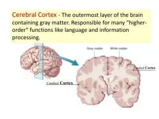
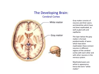
I. Organization General Organization. The cerebral cortex is an overlay (or layer) of gray matter that covers the white matter of the cerebral hemispheres. In the human brain, to increase cortical volume within the skull, both the overlying gray and underlying white matters fold extensively forming gyri (ridges) and sulci (valleys). The word fissure (e.g, lateral, calcaryne) is used sometimes to designate prominent (i.e, deep) sulci.
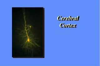
I. Organization Classification of Cortex Based on Cell Layers. Based on how nerve cell bodies and their processes are layered (or stratified), as well as their phylogenetic development, three types of cortex have been distinguished: • Archicortex. Three layered cortex (e.g, hippocampus proper). • Paleocortex. Three-five layered cortex (e.g, olfactory cortex). • Neocortex. Well defined, six-layered cortex (practically most cortex of the hemisphere). The histological arrangement of cells and processes in different cortical areas gave rise to several cytoarchitectonic (cell architecture) maps near the turn of the 20th century, among which Brodmann’s classification of cortical areas is widely used today. Neurocytology of the Cortex. The most common nerve cell type is the pyramidal neuron (PN). They are distributed to all cortical layers but there is a cell size gradient such that the more superficial layers contain small to medium size PNs while the deeper layers contain medium to large PNs. All PNs release glutamate/aspartate (GLU/ASP) from their axon terminals, thus causing excitatory effects on their postsynaptic targets. The axon of PNs projects to: • A near or distant cortical field on the ipsilateral hemisphere (association axon). • A homologous cortical field on the contralateral hemisphere (commissural axon). • A subcortical center (e.g, basal ganglia, brainstem, spinal cord) (projection axon). Interspaced among the PNs the cortex possesses a large variery of different interneurons (short axon neurons such as stellate, basket, etc). Interneurons may be both excitatory (GLU) and inhibitory (GABA)
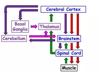
II. Afferent/Efferent Connections Afferents • Afferents from ipsi (associational) or contralateral (commissural) cortex contain GLU and terminate in the upper cortical layers. • Afferents from subcortical modulatory centers such as nucleus basalis (ACh), locus ceruleus (NE), brainstem raphe (ST) and midbrain (DA) end in alllayers. • Afferents from specific thalamic nuclei (contain GLU) terminate in cortical layer 4. Note: Figure on thalamocortical relations in Thalamuslecture illustrates the projection from each of the thalamic nuclei to a distinct cortical field. For example VPM/VPL project to areas 3,1,2; VA to area 6; VL to area 4, etc.
II. Afferent/Efferent……(cont.) Efferents. All originate from from pyramidal neurons. • Short association fibers (U-shaped) interconnect adjacent gyri. • Commissural fibers use the corpus callosum to interconnect homologous cortical areas (e.g, ispsi/contra area 6). • Subcortical projection fibers In the white matter of the hemisphere form the “corona radiata” before passing in the internal capsule to the brainstem and spinal cord. • Long association fibers form the following distinct bundles in the white matter of the hemisphere: - Superior longitudinal fascicle. It interconnects the frontal lobe with the parietal and occipital lobes. Functions in eye movements. - Arcuate fascicle. Interconnects sensory and motor speech areas. - Uncinate fascicle. Interconnects anterior temporal lobe with orbitofrontal gyri. It has limbic functions. - Cingulum Interconnects the cingulate gyrus with parahippocampal gyrus and septal area. Functions in the formation of new memories. - Corpus callosum. Main commissural bundle of the brain. Interconnects most homologous cortical areas in frontal, parietal occipital and superior temporal gyri. - Anterior commissure. Commissural fibers interconnecting the olfactory bulb, olfactory areas and anteriorfd temporal gyri.
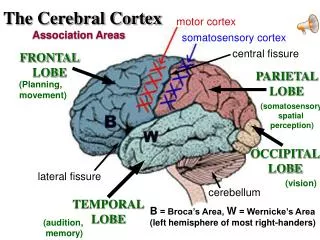
III. Brodmann’s Functional Areas Frontal Lobe Area 4. The primary motor area is found in the posterior part of the precentral gyrus, anterior to the central sulcus. There is a precise representation (motor homonculus) of the contralateral musculature of the body and face in this area. Area 6. The premotor area is anterior to 4 and considered a unimodal association area. The continuation of this area in the medial wall of the hemisphere contains the supplementary motor cortex (SMA). (SMA and Functions in the planning of motor behaviors. Area 8. Anterior to 6 in the middle frontal gyrus it is the frontal eye field. Functions in voluntary conjugate eye movements. Areas 9,10,11. Prefrontal lobe (anterior frontal pole). Multimodal association areas that function as the executive center for the entire brain. Together these areas analyze, judge and plan our actions before putting them into effect. Areas also function to put a “brake” to (or inhibit) our instinctual behaviors. Areas 44, 45 (in most brain on the left hemisphere, dominant for speech). It is known as Broca’s on the pars opercularis (44) and triangularis (45) of the inferior temporal lobe. Controls motor aspects of speech.
III. Brodmann’s……..(cont.) Parietal Lobe Areas 3,1,2. Primary somatosensory area fall in the postcentral gyrus immediately posterior to the central sulcus. There is a precise representation (sensory homonculus) of the contralateral body dermatomes and face area (trigeminal) for pain, temperature, proprioception and vibratory sense sense. Areas 5, 7. Superior parietal lobule. Extend to the medial hemisphere wall where they are known as precuneus. Function in shape recognition (stereognosis) of objects felt with sense of touch. They also functions in visually guided hand behaviors (eye-to-hand co-ordination). Areas 39,40. Area 40 is in the supramarginal gyrus and area 39 in the angular gyrus. Both areas comprise the inferior parietal lobule. In the dominant hemisphere for speech (usually the left) both areas function in complex motor and cognitive functions such as understanding of words and symbols. In the non-dominant hemisphere these areas function to provide spatial perception of contralateral body parts and visual hemifield. Area 19. Visual association area. Overlaps into the occipital lobe
▼ Frontal Lobe 5 4 6 Parieto- occipital Sulcus 7 213 ▼ SFG 8 SM 40 A 39 MFG 19 POp 44 18 17 22 STG PT 45 SFS POrb ▲ MTG IFS ▲ Pre-occipital Notch Lateral Fissure STS ITG MTS