Pulmonary echinococcosis
220 likes | 581 Views
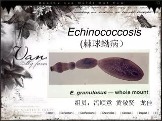
Pulmonary echinococcosis. John-Henry Corbett Department of Radiology University of the Free State 02/2012. Echinoccosis or hydatid disease is caused by larval stage of the echinococcus tapeworm Four species E. granulosis E. multilocularis E. vogeli E. oligarthrus

Pulmonary echinococcosis
E N D
Presentation Transcript
Pulmonary echinococcosis John-Henry Corbett Department of Radiology University of the Free State 02/2012
Echinoccosis or hydatid disease is caused by larval stage of the echinococcus tapeworm • Four species • E. granulosis • E. multilocularis • E. vogeli • E. oligarthrus • Vast majority of infestations in humans are caused by echinococcus granulosis • Worldwide distrubution and concentrated in sheep raising areas
Map shows areas in which hydatid disease is endemic due to the transmission of E granulosus by means of the dog-sheep cycle (solid red areas). Red stripes indicate areas where transmission occurs by means of alternative life cycles in which carnivores such as wolves and foxes serve as definitive hosts and goats, camels, and horses serve as intermediate hosts
Life cycle of E granulosis • Involves two hosts • Dog / carnivore – definitive host • Sheep – intermediate host (most common) • Adult worm of the parasite lives in the proximal small bowel of the definitive host • Eggs are released into the hosts intestine and excreted in feces • Intermediate host ingest ovum while grazing on contaminated ground • Ovum loses its protective layer during digestion in duodenum • Released hexacanth embryo or oncosphere passes through intestinal wall into portal circulation • Develops into a cyst within the liver • Life cycle is completed when definitive host eats the viscera of the intermediate host
Humans may become ‘accidental’ intermediate hosts through • contact with definitive host (dog) • Ingestion of contaminated water or vegetables • Once parasite passes through the intestinal wall to reach portal venous circulation, the liver acts as first line of defense • Therefore the most frequently involved organ (75%) • Lung (15%) • Other locations (10%)
Hydatidcyst structure • Three layers • Pericyst • Outer layer • Composed of inflamed fibrous tissue derived from the host • Form a dense and fibrous protective zone • Ectocyst • Middle layer • Acellular, laminated membrane • Endocyst • Innermost layer • Germinative layer • Gives rise to secondary cysts / brood capsules / daughter cysts
Lungs are the second most common site of hematogenous spread in adults • Most common site in children (25%) • Most cysts acquired during childhood remain asymptomatic • Later diagnosed incidentally at chest radiography • Pulmonary cysts are • Multiple in 30% of cases • Bilateral in 20% of cases • Located in lower lobes 60% of cases • Calcification rare 0,7% • Pulmonary increase in size 1-5cm per year
Sudden coughing attacks, hemoptysis and chest pain are the most common clinical symptoms • After cyst rupture – expectoration of cyst fluid, membranes and scolices may occur • Cyst rupture • Spontaneous • Trauma • Secondary infection • Rupture in pleural cavity may occur • Allergic episodes may occur after cyst rupture, but fatal anaphylaxis is uncommon • Bacterial infection of the cyst afer rupture is the most common serious complication
Radiological • Uncomplicated cysts • Well defined,homogenous, round to oval masses • Surrounded by normal lung tissue • May vary from 1-20cm in size • Cyst shape may vary on inspiratory and expiratory films • (or supine and erect) • Cyst growth causes erosions in the bronchioles that are included in the pericyst • Air is introduced between the pericyst and the laminated membrane • Air collection appears as a thin radiolucent crescent in the upper part of the cyst • Known as the crescent or meniscus sign • Some authors consider this as sign of impending rupture and indication for emergency thoracotomy
Air continues to enter this space between pericyst and ectocyst • Two layers seperate completely • Cyst shrinks and ruptures • Allows for passage of air into the endocyst • Air-fluid level inside the endocyst + air between the pericyst and ectocyst = onion peel appearance • Cumbo sign • After partial expectoration of the endocyst fluid and scolices • Cyst empties • Collapsed membranes can be seen inside the cyst • Serpent sign • When it has completely collapsed • Crumpled endocyst floats freely in the cyst fluid • Water lily sign / Camelotte sign • When all fluid has been expectorated • Remaining solid components will fall to the most dependant part of the cavity • Mass within a cavity/ Monod’s sign
Diagnosis • Made with combination of imaging and serology • Enzyme-linked immunosorbent assay or indirect haemagglutination test is commonly used as an initial screen • positive in only 50% of patients with pulmonary hydatidosis • 90% of patients with hepatic cysts
Treatment • Surgery considered the treatment of choice • Parasite can be completely removed and the patient cured • Options for lung cysts • Lobectomy • Wedge resection • Pericystectomy • Intact endocystectomy • Capitonage • Prevent spill of cyst contents to avoid intraoperative dissemination and recurrance • Delivery of intact cyst • Cyst fluid aspiration ± use of scolicidal solution • Hypertonic saline, povidine iodine, formalin, ethanol, hydrogen peroxide, 1% formalderhyde • Agent must remain in contact with cyst for at least 15 minutes • Pre operative therapy with albendazole • “PAIR” of pulmonary cysts not routinely indicated • Medical therapy starts > 4 days prior to surgery and continues for 3-6 months
References • Morar R, Feldman C. Pulmonary echinococcosis. European Respiratory Journal 2003 ; 21 : 1069-1077 • Pedrosa I, Saiz A, Arrazola J, et al. Hydatid disease : Radiologic and Pathologic Features and Complications. Radiographics 2000 ; 20 : 795-817 • Balikian JP, Mudarris FF. Hydatid disease of the lungs : A roentgenologic study of 50 cases. American Journal of Roentgenology 1974 ; 122 – 4 : 692-707