Download
1 / 51
560 likes | 1.02k Views
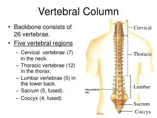
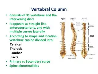
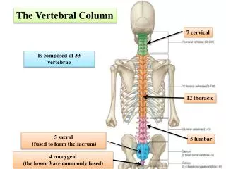
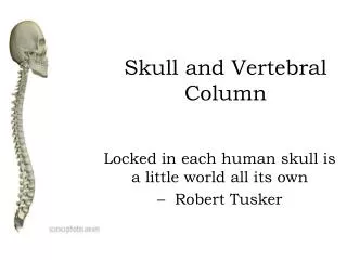
Vertebral Column. The human vertebral column is built up of a series of vertebrae. 7 Cervical, 12 Th., 5 Lumbar, 5 Sacral (Fused as Sacrum), 4 coccygeal (fused as coccyx). Primary Curves: Thoracic and sacral curves - present during fetal development

E N D
The human vertebral column is built up of a series of vertebrae • 7 Cervical, 12 Th., 5 Lumbar, 5 Sacral (Fused as Sacrum), 4 coccygeal (fused as coccyx). • Primary Curves: • Thoracic and sacral curves - present during fetal development • Secondary Curves:-Lumbar and –cervical curves-appear after birth
Typical Vertebrae Body: central strong part. Size increases as we go down towards lumbar region • Superior and inferior surfaces of body (plateaus) • Its post. Surface showing one or more foramina for basivertebral vein. Vertebral Arch: • Pedicles, 2 Laminae. Seven processes: 1) Transverse Processes(2) 2) Spinous Process 3) Facets – superior articular and inferior articular (2) • Vertebral Foramen: • Intervertebral Foramen:
Cervical Vertebrae • Contain 7 vertebrae – C1 – C7 • Smallest vertebrae • Body upper surface is concave • Spinous process – bifid, shorter than diameter of vertebral foramen • Transverse process: Contain transverse foramina • Protect vertebral arteries & veins that go to the brain • Formed of ant. Root, post.root, and costo transverse bar. • Ant. And post. Roots have tubercles, the tubercle of 6th vertebra is called carotid tubercle. • Ant. root.and tubercle ,costotransverse bar represent costal element while the post. root represent the true transverse process.
-Atlas (C1): • articulates with occiptal condyles of skull • has no body or spinous process. -2 lateral masses from which arise transverse processes. • -has a large, round foramen with anterior and posterior arches. • Axis (C2): • supports the atlas -has he dens or odontoid process. • has heavy spinous process • to attach muscles of head and neck • Axis and atlas bodies fuse during development to form the dens
Vertebra prominens (C7): • transitions to thoracic vertebrae • has a long spinous process ,can be felt. • The foramen transverserium for vertebral vein • a traumatic dislocation of cervical vertebrae
The Thoracic Vertebrae • Characteristics : • have heart-shaped bodies • larger bodies than in C1–C7 • smaller vertebral foramen than in C1–C7 • long, slender spinous processes • Dorsolateral surfaces of body have costal facets-which articulate with heads of ribs. • Costal facets is present also on transverse processes to articulate with tubercle of ribs (except 11th and12th ribs)
Has an upper circular complete facet on the body to articulate with 1st rib. • Semilunar lower facet for 2nd rib. - spine thick ,horizontal • T10:has no lower facet on the body. • T11: kidney shaped body like lumbar vertebrae. • -single facet on the body close to upper border • -transverse process has no facet. • T12:single facet away from upper border. • -the transverse process has no facet. • -the inf. Articular processes directed laterally like lumbar vertebrae • T1
The Lumbar Vertebrae Characteristics L1–L4: • largest vertebrae • oval-shaped bodies • thicker bodies than T1–T12 • no costal or transverse costal facets • triangular vertebral foramen • Sup. articular process directed backwards and medially. -mammillary process. • Inf. Art. Process directed laterally. • T. process: long, tapering, has accessory process on post. Surface. -L5:large body. -T. process large bulky Attached to whole side of the pedicle
The Sacrum • Composed of 5 fused sacral vertebrae • Triangular have base and apex. • Anterior concave and post. Convex surfaces. • It has lateral surface and sacral canal. • Articulations: -by the base with 5th lumbar(2nd cartilagenous). -by apex with coccyx.(2nd cartilagenous). -by lateral surface ( auricular surface ) with ilium (sacroiliac joint). • Ant sacral foramina: 4 pairs for ventral rami of upper 4 sacral nerves .+lateral sacral arteries. • Dorsal sacral foramina:4 pairs for dorsal rami of sacral nerves. • Intermediate area between foramina show transverse ridges that indicate fusion of vertebrae.
The Sacrum • Sacral Apex – narrow, inferior portion • Base – broad, superior surface • Sacral Promontory • Important landmark in females – • Used during pelvic exams and during labor & delivery • A prominent bulge @ anterior tip of base • Ala – a wing extending on either side of sacrum
The Sacrum Sacral Canal Ala ofsacrum Auricular Surface Sacral Tuberosity Sacral promontery
-lateral mass: part of sacrum lateral to foramina represents costal element ant. And T. processes post. -Sacral canal • Extends the length of sacrum – nerves & membranes are in this canal -The auricular surface • Thickened, flattened area (on lateral surface) – where articulation w/ pelvic girdle is (sacroiliac or SI joint) -Sacral tuberosity • Site of attachment for ligaments that stabilize the SI joint. -median sacral crest…..fused spines. -intermediate sacral crest……fused articular processes. -lateral sacral crest………. fused T. processes. -sacral hiatus: at lower end of median crest…. Lamina of 5th vertebra failed to fuse leaving a jap. -sacral cornu: free inf. Art. Process of 5th vertebra
Base Sacral Promontory Ala Sacral Apex
Intervertebral Discs -2nd cartilaginous joints. -number 23. -Increase in size from C to L (3mm to 9 mm). -Make up 20-30% of length of column. -the 1st disc between C2 and C3 and the last betweenL5 and S1. - discs are avascular except periphery supplied from adjacent vessels. -disc prolapse commonly occur between L4,5 or between L5 and S1. -PROLAPSE COMMONLY OCCUR IN POSTEROLATERAL DIRECTION WHERE IT IS NOT SUPPORTED BY POST. LONGTUDINAL LIGAMENT.
Two Components • Outer rim of fibrocartilage called the anulus fibrosus. • Connects vertebral bodies in a fibrocartilaginous joint (no capsule, little motion). • Anulus encloses a central mass called the nucleus pulposus. • N.P. About 80-90% water, less with increased age • Contains a mucopolysaccharide matrix • Neither blood vessels or nerves penetrate nucleus
Structure deforms when pressure is put on vertebral column as in weight bearing • Acts as a shock absorber • Annulus totally encloses the nucleus and keeps it under constant pressure • As you get older, the H2O content decreases and the nucleus becomes more fibrocartilaginous, therefore less easily deformable and more easily damaged
Nucleus, when under extreme pressure, can herniate or extrude from the disc in a posterior or posterior-lateral direction • Usually occurs in cervical or lumbar region • Nucleus can put pressure on spinal nerve causing refereed symptoms (motor and sensory) • Can cause pressure on cord itself if true posterior
Major Ligaments of the Spine • Anterior Longitudinal Ligament - ALL • Dense band along anterior and lateral surface of the vertebral bodies from C2 to sacrum • Superficial - bridge several vertebrae • Deep – short, run from V to V, blends with fibers of anulus fibrosus • Limits extension of V column • From C1 to skull, called anterior Atlanto-Occipital Membrane
Major Ligaments • Posterior Longitudinal Ligament • Runs along posterior surface of vertebral bodies (anterior to spinal canal) • C2 to Sacrum • Short fibers attach ligament to posterior disc, reinforce disc posteriorly • Superiorly, continues to occiput, called Tectorial Membrane • Limits flexion
Ligaments • Supraspinous • Spinous process to spinous process – tip to tip • C7 to sacrum • Limits flexion • In cervical region, becomes much thicker with a greater elastic content • Called Ligamentum Nuchae
Ligaments • Interspinous • Found between spinous processes • Most well developed in lumbar region • support
Ligaments • Ligamentum Flavum • Connects lamina of one to lamina of the other • Found from axis to sacrum • Limit flexion • Continuation to the skull is called Posterior Atlanto-Occipital membrane
Ligaments • Intertransverse • Between transverse processes • Limit lateral flexion. • PEDICLE IS HE ONLY PART OF VERTEBRAL ARCH HAVE NO LIGAMENTS TO FORM INTERVERTEBRAL FORAMINA FOR SPINAL NERVES
Sacralization • Where 5th lumbar vertebrae takes on characteristics of the sacrum and may be partially or completely fused with sacrum • Lumbarization:Superior aspect of the sacrum assumes characteristics of the 5th lumbar vertebrae
Atlanto-Axial Joint • Atlas and Axis • Pivot • Two convex superior facets of axis with two concave inferior facets of the atlas • Atlas also posses a facet on the internal surface of the anterior arch which articulates with the dens of the axis • Major ligaments from spine support – Ant. Atlanto-Occipital, Tectorial Membrane, Post. A-O
A-A Joint • Alar – from dens to occiput • Transverse - around dens • Cruciate • Sup. Longitudinal Band • Inferior Longitudinal Band • Transverse
Atlanto-Occipital Joint • Two concave superior facets of atlas articulate with two convex surfaces of occipital condyles of the skull • Supported by major ligaments • Small saddle joint • Very limited motion – nodding type motions in all directions.