Download
1 / 38
410 likes | 756 Views
Amniocentesis and CVS. Dr. Joseph Har-Toov Lis Maternity Hospital Tel-Aviv, Israel. Methods of chromosomal evaluation. Non invasive: Fetal cells from maternal blood preimplantation embryos (PGD) Invasive: amniotic fluid (amniocentesis) placenta (chorionic villus tissue) Fetal blood.

E N D
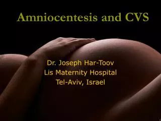
Amniocentesis and CVS Dr. Joseph Har-Toov Lis Maternity Hospital Tel-Aviv, Israel
Methods of chromosomal evaluation • Non invasive: • Fetal cells from maternal blood • preimplantation embryos (PGD) • Invasive: • amniotic fluid (amniocentesis) • placenta (chorionic villus tissue) • Fetal blood
Invasive techniques • Amniocentesis: • Late – second trimester after 15 weeks • Early – earlier than 15 weeks • Chorionic villus sampling (CVS) • Abdominal • Trans cervical • Trans vaginal • Fetal blood sampling
karyotype fish PCR
What can be evaluated? • Chromosomal aberrations: • Trisomy, • Monosomy, • Polyploidy, • Marker chromosome, • Deletion, duplication, inversion, translocation, ring chromosome . • Genetic aberrations (DNA) • Infectious disease • Biochemical markers (AFP)
Amniocentesis • First introduced by Serr and Fuchs and Riis in the 1950s for fetal sex determination • Only at the late 70th a static ultrasound was used to locate the placenta and amniotic fluid pocket • Only In 1983, Jeanty reported a technique of amniocentesis ’’under ultrasound vision’’
Mid Trimester Amniocentesis • Per coetaneous • 20-23g needle • Ultrasound guided • Usually 20cc amniotic fluid • Results – 2 to 3 weeks
complications • Pregnancy loss 0.3-1.0%. • Increase risk: • Needle larger than 18g • Multiple needle insertion • Discoloration of the fluid • High AFP, multiple late abortions, previous vaginal bleeding • Placental perforation – recent studies didn’t find correlation
Complications • Leakage of amniotic fluid (better prognosis than spontaneous leakage) • Amnionitis • Vaginal bleeding • Needle puncture of the fetus • Long term complications: • Respiratory distress?? • Isoimmunization??
Amniocentesis and HIV positive women • Increased rate of vertical transmission • Chemoprophylaxis previous to amniocentesis appears to be beneficial in preventing vertical transmission
Multiple Gestation • Three methods: • Indigo carmine injection to the first sac • A single needle puncture sampling technique (Jeanty 1990) • Simultaneous visualization of two needles on each side of the separating membrane (Bahado-Singh 1992) • Abortion risk – probably higher • Detailed description of fetus position and placental location
Early Amniocentesis: 9-14 weeks • Introduced at late 80th • 10-14 weeks gestation • Only the amniotic (inner) sac should be aspirated • Approximately 1 cc for gestational age • Higher rate of pregnancy loss, talipes equinovarus, and post procedural amniotic fluid leakage • laboratory failure op to 20%
Chorionic villus sampling • Was developed in the 80th • percutaneous transabdominal with 19-20g needle
Chorionic villus sampling • Was developed in the 80th • percutaneous transabdominal • transvaginal • transcervical
15-30mg each aspiration • 20mg ideal for cytogenetic testing • 30-40mg for cytogenetic and other direct molecular and biochemical tests
CVS results • Direct analysis examines the trophoblast cells of the placenta (very rapidly dividing cells) • Results in few hours • greater vulnerability to mitotic error • Cultured analysis examines the fibroblast like cells of the villus stroma or mesenchymal core. • Approximately 7-10 days • Accurately reflect the chromosomes of the fetus.
Risk of invasive procedure • Early amniocentesis: • High pregnancy loss • High fetal malformations • High rate of multiple needle insertions (4.7%) • High rate laboratory failures (1.8%) • Late amniocentesis: • “Low” pregnancy loss (0.3-1%) • Low rate of multiple needle insertions (1.7%) • Low rate laboratory failures (0.2%)
Risk of invasive procedure - CVS • Transabdominal CVS as safe as second trimester amniocentesis • Trans abdominal and transcervical CVS are equally safe and efficacious, provided that centers have expertise with both approaches • In approximately 3–5% of cases, clinical circumstances will support one approach over the other • Limb reduction – not after 9 weeks
mosaicism • True chromosomal mosaicism is when two or more abnormal cells lines are detected in two or more culture flasks from the same individual. • Pseudomosaicism is a term used to describe two abnormal cell lines that are found in only one culture flask (not reported to the patient)
mosaicism • Most often involving trisomic cell and normal cells • 1-2% of pregnancies undergoing CVS • 0.1% of pregnancies undergoing amniocentesis • Clinical outcome of chromosomal mosaicism is strongly dependent on the specific chromosome involved and the number of trisomic cells in both the placenta and the fetus
Mosaicism (trisomic cells) in CVS • Option of an additional prenatal diagnostic procedure (amniocentesis or fetal blood sampling)
Mosaicism (trisomic cells) in CVS • Four possible conditions: • Mosaicism only in the placenta not affecting the fetus or placental function. • Mosaicism only in the placenta not affecting the fetus but alter placental function (IUGR) • Trisomy cells are both in the placenta and in the fetus • Trisomy cells in the placenta and uniparental disomy in the fetus
Mosaicism (trisomic cells) in amniotic fluid • Probably there are trisomic cells in the fetus • The true level and distribution of trisomic cells cannot be accurately assessed with any prenatal procedure • Ultrasound is often the best judge of how a baby is developing
Uniparental Disomy • Arises when an individual inherits two copies of a chromosome pair from one parent and no copy from the other parent • Maternal UPD – two copies from the mother • Paternal UPD – two copies from the father
How does UPD happen? • Loss of a chromosome from a trisomic zygote, "trisomic rescue" • Duplication of a chromosome from a monosomic zygote, "monosomic rescue" • Fertilization of a gamete with two copies of a chromosome by a gamete with no copies of the same chromosome, called gamete complementation.
Trisomic rescue following an error in meiosis heterodisomy
UPD - health concerns in people for two possible reasons: • Parental imprinting in the case of heterodisomy and isodisomy • Unmasking of recessive conditions in some cases of isodisomy
Clinical consequences of UPD • molecular UPD testing should be considered for certain chromosomes (including 6, 7, 11, 14, 15) that are known to have adverse phenotypic imprinting effects.
Factors considered when trying to predict the outcome of mosaicism • the chromosome involved • A mosaic finding 18 or 21 is likely to have worse implications • mosaic finding for trisomy 15 or 16 is likely to have less implications (trisomy 15 or 16 cells cannot survive )
Factors considered when trying to predict the outcome of mosaicism • The tissues affected and level of trisomy in those tissues • The tissue affected cannot be evaluated • The level of trisomy can be only estimated
Factors considered when trying to predict the outcome of mosaicism • method of ascertainment • CVS shows that the placenta is affected • Amniotic fluid suggests that at least one fetal tissue may be affected • Fetal blood sampling confirms the diagnosis of chromosomal mosaicism
Factors considered when trying to predict the outcome of mosaicism • ultrasound findings • presence/absence of uniparental disomy • number of previous case reports known in the literature