Presentation spinal cord
Presentation spinal cord. Spinal cord. Origin: foramen megnum continous with medulla oblongata of brain Termination in adult at the lower boarder of L1 in child at the upper boarder of L3 . Menings. The spinal cord is surrounded by three membranes 1 dura mater 2 :arachnoid mater

Presentation spinal cord
E N D
Presentation Transcript
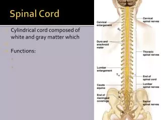
Spinal cord • Origin: • foramen megnum continous with medulla oblongata of brain • Termination • in adult • at the lower boarder of L1 • in child at the upper boarder of L3
Menings The spinal cord is surrounded by three membranes 1dura mater 2 :arachnoid mater 3:pia mater : Function’ Protection Also by cerebrospinal fluid present in the subarachnoid space
In the cervical region • it gives origin to the brachial plexus • lower thoracic region and lumber region • it gives origin to lumbosacral plexus . • superiorly • the spinal cord is fusiformly enlarge the enlargement is referred as the cervical and lumber enlargement • inferiorly • the spinal cord tapers off into the conus medullaris from the apex of which a prolongation of pia mater the filum terminale descend to be attached to the posterior surface of the coccyx.
location • The cord lie in midline • anterior median fissure • posterior median sulcus. • Along the entire length of the spinal cord are attached 31 no of spinal nerves by the • anterior or motor roots and • Posterior root or sensory • posterior root ganglion • cells which gives rise to peripheral and center nerve fibber
Structure of spinal cord • gray mater • inner • white mater • Outer • GRAY MATER • On croos section the gray mater is seenH-shapedpillar with • anteriorcolumn or horns • posterior column or horns • lateral gray column or horn (THORACIC AND LUMBER) • united by • gray commissure • With central canal
Alpha efferent nerve large Multipolar It innervates the skeletal muscle Axon pass out in anterior roots of spinal nerves Gamma efferent Small Multipolar It innervates intrafusal muscle fibers of neuromuscular spindles Axon may pass out in anterior roots of the spinal nerves Nerves cell groups in the anterior gray column
Nerve cell of the anterior gray column is divided into three basic groups • (1) MEDIAL GROUP • (2) CENTRAL GROUP • (3) LATERAL GROUP
Medial group EXTENTION • WHOLE SPINAL CORD • innervate • muscle of • neck, • trunk, • intercostal • abdominal
(2)Central group: EXTENTION cervical, lumber, sacral segments Three nuclei (a) phrenic nucleus (C3’4’5) INERVATE DIAGHPHRAM (b)accessory nucleus) (C5 OR 6) INNERVATION sternocliedomastoid and trapezius muscle (c) lumbosacral nucleus (L2 TO S1) INNERVATION unknwon distribution
Lateral group • Extention • cervical and lumbosacral segment • Innervation • Muscles • (1) upper limb • (2) lower limb
Nerve cells of the posterior gray column • four nerve cell group • 1 substantia gelatinosa • 2 nucleus propius • 3 nucleus dorsalis (clarks column) • 4 visceral afferent nucleus • First two • extention • through out the length of the cord • other two • extention • lumber and thoracic segments
Substantia gelatinosa • location • apex of the posterior gray column • composed • Golgi type 2 neuron • function • receives afferent fiber associated with • pain , • temperature • touch. • Furthermore it receive input from the descending fibers from the supraspinal level .
Nucleus propius • Location • Below s g • Function • senses of • position • movement (proprioception) • two points discrimination • vibration
Nucleus dorsalis • Location • base of the posterior gray column • extending • C8 to L3 4 • FUNCTION • proprioceptive endings neuromuscular spindles and tendon spindle
Visceral afferent nucleus • LOCATION • lateral to the nucleus dorsalis • EXTENTION • T1to L2 • FUNCTION • receiving visceral afferent information
Nerve cell group lateral gray column • Extend • T1 TO S4 • Cells • T1 TO L3 • preganglionic sympathetic nerve fiber • CELLS • S 2,3,4 • preganglionic parasympathetic fiber
The gray commissure and central canal • LOCATION • the anterior and posterior gray columns on each side are connected by a transverse gray commissure so that the gray column r in the central of the gray commissure is situated central canal. • Superiorly • above this it open into the cavity of the fourth ventricle • continuous with the central canal of the caudal half of the medulla oblongata • Inferiorly • It is closed • conus medullaris it expend into the fusiform terminal ventricle • terminate below with in the root of the filum terminale • It is filled with cerebrospinal fluid and is lined with epithelium called the ependyma
IT resembles letter H • posterior gray commissure • The part of the gray commissure that is situated posterior gray canal • Anterior gray commisure • lie anterior to the canal
White mater • It is divided into • anterior lateral • posterior white columns or finiculi. • anterior column • location • lie on each side lie in between the midline and the point of emergence of the anterior nerve root . • lateral column • location • between the emergence of the anterior nerve root and the entry of the posterior nerve root the • posterior column • location • in between the entry of posterior nerve root and midline
Structure • composition • in centrral nervous system the white mater of spinal cord consist of a mixture of • nerve fiber • neuroglia • blood vessel • it surrounds the gray mater • its white color is due to the high proportion of myelinated nerve fiber
Blood Supply of the Spinal Cord The spinal cord receives its arterial supply from three small, longitudinally running arteries: the two posterior spinal arteries and one anterior spinal artery. The posterior spinal arteries, which arise either directly or indirectly from the vertebral arteries, run down the side of the spinal cord, close to the attachments of the posterior spinal nerve roots. The anterior spinal arteries, which arise from the vertebral arteries, unite to form a single artery, which runs down within the anterior median fissure. The posterior and anterior spinal arteries are reinforced by radicular arteries, which enter the vertebral canal through the intervertebral foramina. The veins of the spinal cord drain into the internal vertebral venous plexus.
Injuries in children • Children account for 1-10% of all spinal injuries. • Motor vehicle accidents account for most injuries, followed by falls and sports. • Most serious spinal injuries in children involve the cervical spine.
In children less than 8 years of age, most injuries are between the occiput and C2: • Fulcrum of movement located at C2-3 in children, C5-6 in adults • Significant ligamentous and joint capsule laxity • Relatively large head and weak neck muscles • Horizontal orientation of facet joints • Incomplete ossicification of odontoid process
Injuries in adults
Terminology • Plegia = complete lesion • Paresis = some muscle strength is preserved • Tetraplegia (or quadriplegia) • Injury of the cervical spinal cord • Patient can usually still move his arms using the segments above the injury (e.g., in a C7 injury, the patient can still flex his forearms, using the C5 segment) • Paraplegia • Injury of the thoracic or lumbo-sacral cord, or cauda equina • Hemiplegia • Paralysis of one half of the body • Usually in brain injuries (e.g., stroke)
What are the differences between UMN and LMN? (e.g., cauda equina vs. myelopathy)
Thoracic injuries (T2-L1) • Paraparesis or paraplegia • UMN (upper motor neuron) signs
Cauda equina injuries (L2 or below) • Paraparesis or paraplegia • LMN (lower motor neuron) signs • Thigh flexion is almost always preserved to some degree
What is the difference between cauda equina and conus medullaris syndrome?