Download
1 / 25
270 likes | 429 Views
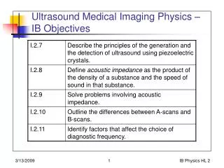
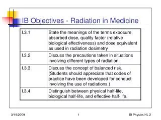
IB Objectives - Radiation in Medicine. IB Objectives - Radiation in Medicine. Radiological Medicine. Terminology Activity Source-related Exposure Amount of ionization Dose Amount of energy absorped Equivalent dose Biological effects. Image from: reactor.reed.edu/pictures.html.

E N D
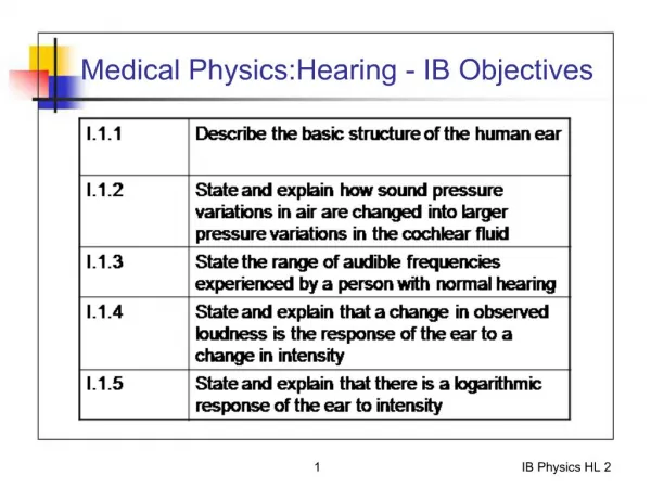
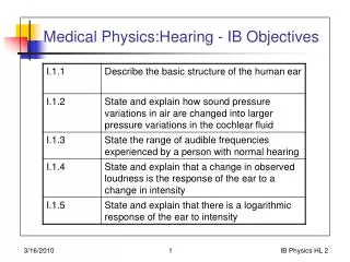
IB Objectives - Radiation in Medicine IB Physics HL 2
IB Objectives - Radiation in Medicine IB Physics HL 2
Radiological Medicine • Terminology • Activity • Source-related • Exposure • Amount of ionization • Dose • Amount of energy absorped • Equivalent dose • Biological effects IB Physics HL 2 Image from: reactor.reed.edu/pictures.html
Activity • Number of decays per unit time • Depends on atomic type and amount • Other equations: • What is the activity of 5 g of 131I (t1/2 of 8 days) IB Physics HL 2
Exposure • Measures amount of ionizing radiation something is “exposed” to • Ionization -> charge separation • Taken relative to charge separation in air • ~ Charge separation in tissue • For X-rays and -rays IB Physics HL 2 Image from:www.rogerwendell.com/nukes.html
Absorbed Dose • Measures how much radioactive energy actually deposited in an object (relative to mass) • In air, 34 eV needed to create one ion pair, so • 1 C/kg in air is34 eV x 1.6 x 10-19 J x 6.25 x 1018 electrons = 34 J/kg= 34 Gy • Other materials would differ (energy to create pair of ions) IB Physics HL 2
Equivalent Dose • Absorbed dose does not relate the biological effect of specific radioactive decay particle. • Equivalent dose does take biological “effectiveness” into account • H (equivalent dose) (seivert) = Q D(dose) Nwhere Q is Quality FactorD is absorbed dose (Gy)N is other factors (set to 1) • Q = 1 for X-rays, -rays (at 250 keV), protons = 5 for thermal (slow) neutrons = 10 for fast neutrons, particles = 20 for recoiling nuclei IB Physics HL 2
Quality Factor • Also called Relative Biological Effectivenessrelative to 250 keV X-ray IB Physics HL 2
Activity to Equivalent Dose Activity(Bq - Disintigrations/s) Geometric factorsTime factors Qualityfactor Exposure (C/kg) Dose (Gy) Equivalent Dose (Sv) IB Physics HL 2
Radiation Effects on Humans • X-ray, -ray less harmful than electrons, particles • Differing effects on different cells • Reproductive cells very radiation sensitive • Nerve cells not so sensitive • With increasing exposure: • Topical burns • Nausea, diarrhea, fever (radiation sickness) • Loss of hair • Cancer, leukemia • Death IB Physics HL 2
Radiation Effects on Humans IB Physics HL 2
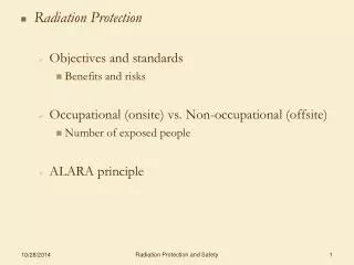
Precautions and Risks • Patient • Practitioner IB Physics HL 2
Precautions and Risks • Patient • Monitor exposure time carefully • Use only procedures that convey net benefit • Keep exposures as low as reasonably achievable • Do not exceed recommended limits for dose • Lead aprons (reduce stray radiation) IB Physics HL 2
Precautions and Risks • Practitioner • Procedures to limit or minimize risk of contamination or exposure • Monitor radiation exposure (film badge) • , -ray, X-ray, and neutron monitoring IB Physics HL 2
Precautions and Risks • Practitioner procedures to minimize risk: • Use lab coat in locations where radioactive material used, handled, or stored • Use disposable gloves • Monitor hands before and after leaving work area • No eating, drinking, or smoking in work area • Clearly label radioactive material IB Physics HL 2
Precautions and Risks • Shielding (patient and practitioner) • Distance (1/R2 fall-off) • Lead, concrete, water: X-rays and -rays • Neutrons: mass (lead, steel) • Lead aprons (patient) IB Physics HL 2
Half Lives • Radiological half-life (physical half-life): TR • Time for half of radioactive isotope to decay • Biological half-life: TB • Time for the body to get rid of half of the radioactive isotope • Effective half-life: TE • Effective half-life of isotope, including both radiological and biological effects • 1/TE = 1/TR + 1/TB orE = R + B (decay constants) IB Physics HL 2
Half Life Example • Thallium-200 has a radiological half-life of 26 hours, and a biological half-life of 42 hours. • What is its effective half-life? • How long will it take for its activity to fall to 1/10th its initial value? IB Physics HL 2
Radiation Treatment of Cancer • Laws of Bergonie and Tribondeau regarding sensitivity of cells to ionizing radiation • More sensitive when cells are: • Young • Simple • High metabolism • Dividing rapidly • These characteristics make cancer cells more susceptible to radiation damage than normal cells IB Physics HL 2
Radiation Treatments of Cancer • Three main classifications • Internal radiotherapyRadioisotope is in body, and becomes localized in affected organ (e.g., I-131 and thyroid cancer) • External radiotherapyRadioactivity from source outside body (e.g., accelerator or radioactive source) • BrachytherapyWhere radioactive source implanted in body near locale to receive radiation IB Physics HL 2
Internal Radiotherapy • Use isotopes which emit -rays, -particles (electrons) • Deposit energy close to where radioisotopes are • Examples • I-131 and thyroid treatment • Yt-90 - liver cancer • P-32 - bone marrow • Sm-153 and breast and prostrate cancers IB Physics HL 2
External Radiotherapy (Teletherapy) • Collimate and shape beams to illuminate target tumor • Reduce illumination of healthy tissue • Beam from accelerator or radioactive source (e.g., Co-60) IB Physics HL 2
Brachytherapy • Use , -emitters to localize energy deposition • Radioactive source is local • Reduce illumination of healthy tissue • Place catheters for placement of wire • Example: Ir-192 and breast cancer, mouth cancer IB Physics HL 2
Radioactive Tracers (Diagnostics) • Use to determine functioning of physiological processes, location • Usually use -emitter • Emerge from body easily • Can be detected by scintillation camera • Want short radiological half-life (<~ day) • Examples: • Cr-51 and bleeding • Sr-90 and bones • Rb-86 and muscles • Tc-99 and multiple organ imaging IB Physics HL 2
Key Ideas for Radiological Medicine IB Physics HL 2