Download
1 / 1
10 likes | 165 Views
Using Solid State NMR to Understand the Molecular Structure of Designer Self-Assembling Proteins MAX8 and RADA16.

E N D
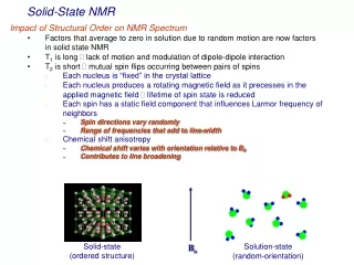
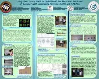
Using Solid State NMR to Understand the Molecular Structure of Designer Self-Assembling Proteins MAX8 and RADA16 Dr. Anant K. Paravastu Ashley Cormier Megan Crombie Kathy Gibbons-Adams Chemical & Biomedical Engineering Graduate Researcher Riversink Elementary School Lakeview Elementary School FSU-FAMU College of Engineering FSU-FAMU College of Engineering Crawfordville, FL 32327 Miami, FL 33167 Tallahassee, FL 32303 Tallahassee, FL 32303 Introduction Proteins are a necessary part of everyday life. Nerves, tissues, and bones all contain proteins we need to survive. Our bodies also make different proteins that help our cells grow, fight infection, and repair injuries. The scientists we worked with this summer were interested in studying designer proteins that were synthetically produced by man. Though these proteins are not as complex as ones that occur naturally, they are important because they can be used for repair and regeneration of cells and nerves. Our immune systems make complicated T-cell proteins to fight diseases. Conclusion and Future Research At this time there is a computerized molecular model of MAX8 in the process of being built and confirmed. Future NMR experiments with RADA16 must continue to be conducted and data must be collected and analyzed to learn more about how the molecules in the protein interact at different temperatures and in different solvents. This will help scientists learn more about the nature of the protein, which will aid in determining the molecular structure. Nuclear Magnetic Resonance (NMR) Next the protein samples are lowered into the middle of the magnet. NMR spectrometry uses a spectrometer that sends radio waves via wires to a magnet. Our magnet generates 11.74 Tesla; the magnetic field of the earth ranges from 0.3-0.6 Gauss and 10,000 Gauss = 1 Tesla. The solid sample spins 25,000 times per second to create a magic angle spinning environment. This allows for the most accurate data collection as samples oscillate between ground and excited states. During spinning Nitrogen gas or compressed air is used to cool the magnet. The protein’s nuclei and electrons emit different radio waves that are recorded through Fourier’s Transform. Megan and Kathy can stand near the NMR magnet because it has an “ultrashielded,” tight magnetic field. Classroom Applications The most important part of this experience was finding a way to take what we learned back to our elementary classrooms. We used beads to represent amino acids that self-assembled into proteins. This is a hands-on way to show students functions that occur for cell and tissue growth. The strands must have the correct components, be in a specific order, and be close enough to each other for self-assembly to occur. The same process is followed in our bodies and with designer proteins. Purpose Our summer mission was to analyze data from NMR spectrometry to better understand the molecular structures of the designer proteins MAX8 and RADA16. This would let scientists use computer programs to create 3-D models of the proteins. Even though scientists are able to synthesize and experiment with these proteins, they are still not completely certain about all of their structural characteristics. Once they understand more about MAX8 and RADA16, they hypothesize that they will be able to use these proteins to deliver medicine. These proteins are particularly interesting because they can self-assemble and then reassemble in new environments. Real world application of these proteins would allow someone to receive an injection of MAX8 or RADA16 to assist in nerve regeneration. A A NMR uses radio waves; the lower frequency allows for detailed information on molecular structure and environment. E B C D These beads represent amino acids. Some will self-assemble to form proteins, some will not. Strand A did not self-assemble; B-E did. B is folded, C has coils and folds, D is coiled, and E has large folds. Protein Synthesis Before we could use NMR spectrometry to analyze data about the proteins, we worked with Dr. Paravastu’s graduate students to synthesize MAX8 and RADA16. In this process scientists can make designer proteins by using specific amounts of amino acids. The amino acids have to follow a particular sequence for each desired protein. Our bodies do this naturally through coding that we are born with in our DNA. When we assisted in making RADA16, first we weighed specific amounts of each amino acid (arginine, alanine, and aspartic acid). They were placed in order on the peptide synthesizer, which injected necessary amounts solvents that helped of each amino acids form the protein. This process takes about an hour per amino acid, which means a gram of RADA16 takes about 16 hours to make. After this, the protein must be cleaved and purified. The end result is about .25g of RADA16 which is worth $1,000.00. • References • Cormier, A. Paravastu, “Solid state NMR structural analysis of designer self assembling proteins,” MRS Conference Poster, Spring (2010). • A. Paravastu, “Optical polarization of nuclear spins in gallium arsenide,” University of California, Berkeley (2004). • A. Paravastu, R. Leapman, W. Yau, R. Tycko, “Molecular structural basis for polymorphism in Alzeimer’sβ-amyloid fibrils,” Proceedings of the National Academy of Sciences of the United States Journal (2008). • H. Yokoi, T. Kinoshita, S. Zhang, “Dynamic reassembly of peptide RADA16 nanofiber scaffold,” Proceedings of the National Academy of Sciences of the United States Journal (2004). • Acknowledgements • Because Dr. Paravastu collaborates with teams at FSU, FAMU, the College of Engineering and the Magnet Lab, we had the unique opportunity to observe many different and diverse scientists throughout the summer. We would like to thank UmeshGoli, Dr. Rufina Alamo, Dr. Derek Lovingood, Dr. Ongi Englander, Dr. KarunyaKandimalla, Dr. Subramanian Ramakrishnan, Dr. Geoffrey Strouse and Dr. Penny Gilmer for helping us experience scientific research on a daily basis. In addition, we appreciated the willingness of Dr. Paravastu’s students Serena Huang, Stefanny Cortes, Alexa Buchannan, and Andres Gutierrez as they allowed us to spend the summer learning with them. Thank you to Dr. Pat Dixon, Jose Sanchez, Carlos Villa, Patrick Enderle, Nguyen Nguyenand the RET participants for their support at the Mag Lab education department; our days with the scientists became more meaningful as we worked together to figure out how to connect authentic research to inquiry-based learning in our classrooms. Our level of understanding would not have been possible without graduate student Ashley Cormier patiently explaining data to us as she worked through the scientific method each day. Finally, we are grateful for everything that Dr. AnantParavastu allowed us to experience as we shared the summer with a true scientist. We will take his undying curiosity about the world back to our classrooms as we try to inspire future generations of scientists. Fourier’s Transform uses mathematical equations to turn radio waves into usable data. Cell phones and computers use it everyday to process information. Megan tunes the probe to find signals for specific elements in RADA16. Challenges with MAX8 and RADA16 Determining the molecular structure of the proteins is difficult because many variables are involved. The NMR graph below shows RADA16 emits different signals when it is heated. There are also concerns about exposure to water vapor in the air and the protein breaking down over time. MAX8 will currently dissolve in a solvent but RADA16 will not, which interferes with the HPLC purification process for proteins. Both proteins seem to form β-sheets and it is possible that RADA16’s β-sheets are keeping it from dissolving. Kathy weighs the delicate powder form of arginine while Serena keeps a careful eye on the digital reading. RADA16 is made with 16 amino acids that follow the RADA pattern; MAX8 is made with 20 amino acids. Coil? Yellow-2.5 mm Probe; 25 kHz MAS; RADA16 4/24/10 no HPLC at room temperature. Red-2.5 mm Probe; 25 kHz MAS; RADA16 4/24/10 no HPLC, CPMAS at 80 C, spinning with N2 gas, a few hours later. Blue-2.5 mm Probe; 25 kHz MAS; RADA16 4/24/10, no HPLC, CPMAS at room temperature, spinning with N2 gas, the next morning. The brown portion of the graph shows the absorption rates of each amino acid during the decoupling process; scientists infer there may be a coil in RADA16’s structure. • RADA16 self-assembles and forms β-sheets that seem to make it insoluble. • MAX8 self-assembles and forms • hairpin β-sheets but it will still • dissolve in different solvents. The peptide synthesizer is ready to go!