Download
1 / 103
1.13k likes | 2.14k Views
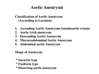
Aortic aneurysm. Dr. Aidah Abu Elsoud Alkaissi An-Najah National University Nursing College. Thorasic Aorta Aneurysm. May be a symptomatic Back, neck or substernal pain Dyspnea, stridor or brassy cough if pressing on trachea

E N D
Aortic aneurysm Dr. Aidah Abu Elsoud Alkaissi An-Najah National University Nursing College
Thorasic Aorta Aneurysm • May be a symptomatic • Back, neck or substernal pain • Dyspnea, stridor or brassy cough if pressing on trachea • Hoarseness and dysphagia if pressing on esophagus or laryngeal nerve • Edema of the face and neck • Distended neck vein • Complications: rupture and hemorrhage
Abdominal Aorta Aneurysm • Client´s awareness of a pulsating mass in the abdomen, with or withourt pain, followed by abdominal pain and back pain • Flang pain or groin pain may be experienced because of increasing pressure on other structures sometimes mottling (the act of coloring with areas of different shades) of the extrimities or distal emboli in the feet alert the clinician to a source in the abdomen
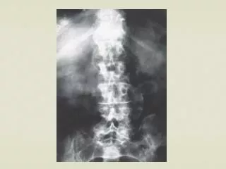
Abdominal Aorta Aneurysm • Pulsating abdominal mass • Aortic calcification noted on x-ray • Mild to severe midabdominal or lumbar back pain • Cool, cyanotic extrimities if iliac arteries are involved • Claudication (ischemic pain with exercise, relieved by rest) • Complication: peripheral emboli to lower extrimities • Rupture and hemorrge
Medication • Thorasic aorta aneurysms are treated with long-term beta blocker therapy and additional antihypertensive drugs as needed to control heart rate and blood pressure • Clients with aortic disection are intially treated with intravenous beta blocker such as propranolol (Inderal) , metoprolol (Lopressor), labetalol (Normodyne) , or esmolol (Brevibloc) to reduce the heart rate to about 60BPM. • Sodium nitroprusside (Nipride) infusion is started concurrently to reduce the systolic pressure to 120 mmHg or less
Medication • Calcium channel blockers also may be used • Direct vasodilator such as diazoxide (Hyperstat) and hydralazine (Apresoline) are avoided as they may actually worsen the dissection • Constant monitoring of vital signs, hemodynamic pressures (via Swan-Ganz catheter and urin outpt are vital to ensure adequate perfusion of vital organs • Following surgical correction of an aneurym, anticoagulant tharapy may be intiated • Heparin therapy is used initially, with conversion to oral anticoagulation prior to discharge • Many clients are manifested indefinitely on anticoagulation therapy, others may use lifelong, low- dose-aspirin therapy to reduce the risk of clot formation
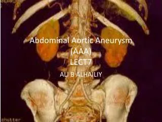
Abdominal Aortic Aneurysm Repair • (Abdominal Aneurysm - Open Repair, AAA Repair, Triple A Repair, Abdominal Aneurysmectomy, Endovascular Aneurysm Repair, EVAR)
Procedure OverviewWhat is an abdominal aortic aneurysm repair? • Abdominal aortic aneurysm (AAA) repair is a procedure used to treat an aneurysm (abnormal enlargement) of the abdominal aorta. • Repair of an abdominal aortic aneurysm may be performed surgically through an open incision or in a minimally-invasive procedure called endovascular aneurysm repair (EVAR).
Procedure OverviewWhat is an abdominal aortic aneurysm repair? • Aggressive control of the blood pressure and prolonged bed restis the usual initial treatment for patients with uncomplicateddissection sparing the ascending aorta (Stanford type B) • asemergency surgery to the descending thoracic aorta carries asubstantial mortality when compared with medical treatment. • Surgery should be reconsidered if there is evidence of aorticrupture, proximal extension of the dissection, or ischaemiccomplications.
The optimal management of patients with suspected dissectionrequires close liaison between district hospitals and cardiacsurgical centres and use of local guidelines for investigationthat reflects the available skill. • Patients with a low clinicallikelihood of dissection who are in a stable cardiovascularstate should undergo prompt local investigation with a nominatednon-invasive technique. • Unstable patients with a high likelihoodof dissection should receive medical treatment and be transferredimmediately to the surgical centre for both diagnostic imagingand management.
If skill in transthoracic or transoesophagealechocardiography is available locally these procedures can beperformed while transport is awaited, but doing so should notdelay transfer. • A videotaped record of the study should accompanythe patient to the surgical centre, where repeat transoesophagealechocardiography can be performed in the anaesthetic room ifnecessary. • This approach minimises delay, an essential stepin lowering the mortality of acute dissection.
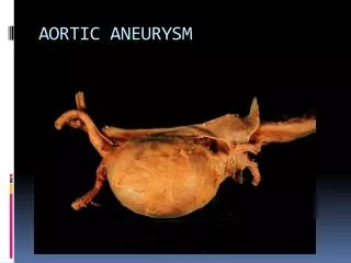
What is an abdominal aortic aneurysm? • An abdominal aortic aneurysm, also called AAA or triple A, is a bulging, weakened area in the wall of the aorta (the largest artery in the body) resulting in an abnormal widening or ballooning greater than 50 percent of the normal diameter (width).
The most common location of arterial aneurysm formation is the abdominal aorta, specifically, the segment of the abdominal aorta below the kidneys. • An abdominal aneurysm located below the kidneys is called an infrarenal aneurysm. An aneurysm can be characterized by its location, shape, and cause. • The shape of an aneurysm is described as being fusiform or saccular, which helps to identify a true aneurysm. • The more common fusiform-shaped aneurysm bulges or balloons out on all sides of the aorta. A saccular-shaped aneurysm bulges or balloons out only on one side.
A pseudoaneurysm, or false aneurysm, is an enlargement of only the outer layer of the blood vessel wall. • A false aneurysm may be the result of a prior surgery or trauma. • Sometimes, a tear can occur on the inside layer of the vessel resulting in blood filling in between the layers of the blood vessel wall, creating a pseudoaneurysm
The aorta is under constant pressure as blood is ejected from the heart. With each heart beat, the walls of the aorta distend (expand) and then recoil (spring back), exerting continual pressure or stress on the already weakened aneurysm wall. • Therefore, there is a potential for rupture (bursting) or dissection (separation of the layers of the aortic wall) of the aorta, which may cause life-threatening hemorrhage (uncontrolled bleeding) and, potentially, death.
The larger the aneurysm becomes, the greater the risk of rupture. • Because an aneurysm may continue to increase in size, along with progressive weakening of the artery wall, surgical intervention may be needed. • Preventing rupture of an aneurysm is one of the goals of therapy
Types of abdominal aneurysm repair: • There are two approaches to abdominal aortic aneurysm repair. • The standard surgical procedure for AAA repair is called the open repair. • A newer procedure is the endovascular aneurysm repair (EVAR).
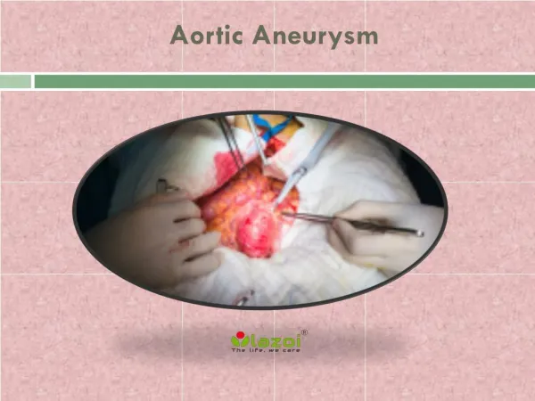
abdominal aortic aneurysm open repair:Open repair of an abdominal aortic aneurysm involves an incision of the abdomen to directly visualize the aortic aneurysm. • The procedure is performed in an operating room under general anesthesia. • The surgeon will make an incision in the abdomen either lengthwise from below the breastbone to just below the navel or across the abdomen and down the center. • Once the abdomen is opened, the aneurysm will be repaired by the use of a long cylinder-like tube called a graft.
The aneurysm is exposed, the aorta is clamped just above and below the aneurysm to stop the flow of blood, the aneurysm is opened and a Dacron graft is placed within the anuerysm • The aneurysm sac is then wrapped around the graft to protect it
Grafts are made of various materials, such as Dacron (textile polyester synthetic graft) or polytetrafluoroethylene (PTFE, a non-textile synthetic graft). • The graft is sutured to the aorta connecting one end of the aorta at the site of the aneurysm to the other end of the aorta. • Open repair remains the standard procedure for an abdominal aortic aneurysm repair.
Complications • Caused by underlying coronary artery disease and chronic obstructive pulmonary duisease • These conditions decreased metabolism of anesthetic, increase the risk of postoperative atelectasis and decrease the client´s tolerance of hemodynamic changes from blood loss and fluid shifts
Complications • To reduce the risk of acute myocardial infarction, one of the most serious complications, clients may undergo coronary artery bypass before aneurysm repair • Prerenal failure can develop for several reasons • The kidney can sustain ischemia from decreased aortic blood flow, decreased cardiac output, emboli, inadequate hydration or the need for clamps on the aorta above the renal arteries during surgery
Complications • Emboli can also develop and lodge in the arteries of the lower extrimities or mesentery • Clinical manifestations include those of acute occlusion in the leg • Bowel necrosis is exhibited as fever, leukocytosis, ileus, diarrhea and abdominal pain • The spinal cord can also cbecome ischemic, resulting in paraplegia, rectal and urinary incontinence or loss of pain and temperature sensation • Spinal cord ischemia tends to occur more commonly when an abdominal aortic aneurysm has ruptured
Complications • Changes in sexual function may also develop following repair of an abdominal aortic aneurysm • Retrograde ejaculation occurs in about two third of male clients and loss of potency occurs in one third of males who have undergo repair of abdominal aortic aneurysm
endovascular aneurysm repair (EVAR)EVAR is a minimally-invasive (without a large abdominal incision) procedure performed to repair an abdominal aortic aneurysm. • EVAR may be performed in an operating room, radiology department, or a catheterization laboratory. • The physician may use general anesthesia or regional anesthesia (epidural or spinal anesthesia).
The physician will make a small incision in each groin to visualize the femoral arteries in each leg. • With the use of special endovascular instruments, along with x-ray images for guidance, a stent-graft will be inserted through the femoral artery and advanced up into the aorta to the site of the aneurysm.
A stent-graft is a long cylinder-like tube made of a thin metal framework (stent), while the graft portion is made of various materials such as Dacron or polytetrafluoroethylene (PTFE) and may cover the stent. • The stent helps to hold the graft in place. • The stent-graft is inserted into the aorta in a collapsed position and placed at the aneurysm site. • Once in place, the stent-graft will be expanded (in a spring-like fashion), attaching to the wall of the aorta to support the wall of the aorta. • The aneurysm will eventually shrink down onto the stent-graft.
Reasons for the Procedure • Reasons an abdominal aortic aneurysm repair may be performed include, but are not limited to, the following: • to prevent the risk of rupture • to relieve symptoms • to restore a good blood flow • size of aneurysm greater than 5 centimeters in diameter (about two inches) • growth rate of aneurysm of more than 0.5 centimeter (about 0.2 inch) over one year • when risk of rupture outweighs the risk of surgery • emergency life-threatening hemorrhage (uncontrolled bleeding) .
Risks of the Procedure • As with any surgical procedure, complications can occur. Some possible complications may include, but are not limited to, the following: • open repair: • myocardial infarction (heart attack) • irregular heart rhythms (arrhythmias) • bleeding during or after surgery • injury to the bowel (intestines) • limb ischemia (loss of blood flow to legs/ feet) • embolus (clot) to other parts of the body • infection of the graft • lung problems • kidney damage • spinal cord injury
EVAR: • damage to surrounding blood vessels, organs, or other structures by instruments • kidney damage • limb ischemia (loss of blood flow to leg/feet) from clots • groin wound infection • groin hematoma (large blood-filled bruise) • bleeding • endoleak (continual leaking of blood out of the graft and into the aneurysm sac with potential rupture) • spinal cord injury • Patients who are allergic to or sensitive to medications, contrast dyes, iodine, shellfish, or latex should notify their physician.
Nursing Care of the client having surgery of aortaPreoperative Care • Preoperative assessment must include detection of concurrent coronary artery disease and cerebrovascular disease • Assess all peripheral pulses for baseline comparison postoperativelyIf emergent surgery is required, time for preoperative care and teaching may be limited • Implement measures to reduce fear and anxiety: • Orient to the intensive care unit, if appropriate • Describe and explain the reason for all equipment and tubes, sucgh as cardiac monitors, ventilators, nasogastric tubes, urinary catheters, intravenous lines and fluids and intra-arterial lines • Explain what to expect following surgery (sights, sounds, frequency of taking vital signs, dressing, pain relief measures, communication strategies)
Nursing Care of the client having surgery of aortaPreoperative Care • Allow time for questions and expression of fears and concerns • These explanation provide a sense of control for the client and family
Nursing Assessment • A thorough nursing history and physical asessment should be performed, because atherosclerosis is a systemic desease process • It is important for the nurse to watch for signs of cardiac, pulmonary, cerebral and lower extrimity vascular problems • The patient should be monitored for indications of rupture of the aneurysm such as diaphoresis, paleness, weakness, tachycardia, hypotension, abdominal, back, groin or periumbilcal pain, changes in sensorium or a pulsating abdominal mass • Attention to the character and quality of the peripheral pulses and the neurologic status • Pedal pulse sites (dorsalis pedis and posterial tibial) and skin lesions on the lower extrimities should be marked and documented before surgery
planning • The overall goals for a patient undergoing aortic surgery include • 1. Normal tissue perfucsion • 2. Intact motor and sensory function • No complications related to surgical repair such as thrombosis or infection
Nursing ImplementationHealth Promotion • The nurse must be aware of cardiovascular disease risk factors and be alert for opportunities to teach health promotion measures to patients in the hospital and the community • Special attention should be given to the patient with a strong family history of aneurysm or any evidence of other cardiovascular disease
Nursing ImplementationHealth Promotion • The patient should be encouraged to reduce risk factors known to be associated with atherosclerosis • These should include controlling hypertension, smoking cessation, and following a diet low in fats and cholesterol • These measures are also done to ensure contiued graft patency following surgical repair
Nursing ImplementationAcute intervention • Preoperative teaching should include a brief explanation of the disease process, the planned surgical procedures, preoperative routines, what to expect immediately after surgery ” e.g., recovery room, tubes/drains” and usual postoperative timelines • Preoperative routine, bowel preparation (laxatives, enemas) and have a preoperative shower with an antimicrobial soap the day before surgery, receive nothing by mouth after midnight the day before surgery and often are given preoperative intravenous antibiotics immediately before surgery • A tour of the ICU before surgery may be of interest to the patient and family
Nursing ImplementationAcute intervention • When the pat arrives in the ICU, an endotrachial tube, an arterial line, a central venous catheter, or pulmonary artery catheter, peripheral i.v line, an indwelling urinary catheter and a nasogastric tube will likely be in place with continous ECG and pulse oximetry monitoring • If the thorax is entered during surgery, chest tube will also be in place, pain medication may be administered via epidural catheter or patient controlled analgesia • Maintaining adequate respiratory function, fluid and electrolytes balance , pain control
Nursing ImplementationAcute intervention • The nurse must monitor graft patency, and renal perfusion • The nurse can also assist in preventing arrhythmia, infections and neurologic complications
Nursing Implementationgraft patency • Maintain adequate blood pressure to promote graft patency. Prolonged hypotention may result in graft thrombosis due to decreased blood flow • Administration of of i.v. Fluids and blood components as indicatedis essential to maintaining adequate blood flow to the graft • Central venous pressure readings or pulmonary artery pressures and urinary output should be monitored hourly in the immediate postoperative period to help assess the patient´s state of hydration
Nursing Implementationgraft patency • Severe hypertention may cause undue stress on the arterial anastomosis • Resulting in leakage blood or rupture at the suture lines • Drug therapy with duiretics or i.v antihypertensive agents may be indicated if severe hypertension persists
Nursing ImplementationCardiovascular status • In individuals with preexisiting coronary artery disease, myocardial ischemia or infarction may occur in the perioperative period due to decreased oxygen supply to the heart or increased oxygen demands on the heart. • Cardiac rhythmias also may occur due to electrolyte imbalances, hypoxemia, hypothermia or myocardial ischemia
Nursing interventions include continous ECG monitoring, frequent electrolyte and blood gas (ABG) determinations, administrations of oxygen and Antiarrhythmic medications as needed • Replacement of electrolytes as indicated, adequate pain control and resumption of preoperative cardiac medications
Nursing ImplementationInfection • The development of a prosthetic vascular graft infection is relatively rare but possibly life threatening complications • Nursing prevention to prevent infection should include ensuring that the patients receives a broad spectrum antibiotic as prescribed • Assess body temperature regularly and report any elevations • Laboratory data should be monitored for elevated WBC • The nurse should ensure adequate nutrition and observe the surgical incision for any evidence of delaying healing, signs of infection or prolonged drainage
Nursing ImplementationInfection • All i.v, arterial and central venous catheter insertion sites should be cared for carefully with the use of sterile technique because they are frequently a portal of antry for bacteria • Meticulous perinial care for the patient withan indwelling urinary catheter is essential to minimize the risk of urinary tract infection • Surgical inncisions should be kept clean and dry