Download
1 / 39
470 likes | 1.07k Views
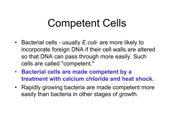
9. seminar/practice. Methods to measure f unctional of the immun o competent cells blast transformation (LPS and ConA activation), polyclonal B and T lymphocyte activation, ELISPOT. Measuring the functional activity of T and B lymphocytes. Topics :.

E N D
9. seminar/practice Methods to measure functional of the immunocompetent cells blast transformation (LPS and ConA activation), polyclonal B and Tlymphocyte activation, ELISPOT
Measuring the functional activity of T and Blymphocytes Topics: Polyclonal activation of T and Bcells via non-antigen-specific stimulation lectin-induced activation α-IgM, α-CD3 or α-TCR antibody allogeneicTcell activation (examination of the immediate-early activation events) Characterization of responses by activated T and B cells activation markers proliferative response: 3H-thymidine incorporation CFSE fluorescence decrease cell cycle events Antibody or cytokine production (ELISA, CBA) Determination of the number of activated T and B cells after the administration of the antigen ELISPOT, Intracellular cytokine staining Pentamer (or tetramer) technics
(review) Phases of the humoral immune response
(review) Phases of T cell response
BCR signaling (review)
TCR signaling (review)
Immunodeficiencies mainly characterized by different functional immunoassays Lymphocyte activation by specific antigen is hardly detected, because of the low number of the antigen specific cells Lymphocyte function can be investigated by polyclonal T/B-lymphocyte activator materials
T cell B cell (mouse) T cell TLR4 (PMA activates protein kinase C) Polyclonal activation of lymphocites by LPS, lectins, PMA/ionomycin BCR- or TCR-specific antibodies may also activate the lymphocytes
Polyclonal B cell activators Activator T cell dependency Ig secretion Human B cells PWM (pokeweed mitogen) no yes SpA (superantigen, staphylococcus protein A) no yes EBV (transforming effect) yes yes Anti-Ig yes In the presence of cytokines Mouse B cells LPS no yes PWM yes yes PPD (purified protein derivate, mycobacterium) no yes Anti-Ig no In the presence of cytokines
Pokeweed (PWM) (Phytolacca americana) – formerly used for colouring red wine (toxic: triterpene saponin) Chenopodiales Phytolaccaceae
Phytohaemagglutinin (PHA) Canavalia ensiformis – Jack-bean, Sword bean
Receptor crosslinking (immediate) phosphorilation steps (seconds-minutes) - Western blot - Bead array Antigen receptors (TCR, BCR), and different other receptors (e.g. cytokine receptors) ic Ca2+ increase - FACS, microscopy Gene activation - RT-PCR Cytokine synthesis - IC cytometry Cytokine secretion • ELISA, ELISPOT - DNA content - IN antigens Cell-cycle/apoptosis Lymphocyte activation Cell division - 3H-thymidine, CFSE, MTT The examination often requires specific Ag-Ab reactions
Western blot It can detect the presence or even phosphorylationstate of specific proteins The cells’ activation stage can be „frozen” at different times, so the eventsof the activation can be monitored in parallel samples. at least 105-106 cells required
Investigation of the presence or absence of Bruton’s tyrosine kinase (BTK) by Western blot X-linked agammaglobulinemia. XLA patients do not generate mature B cells, which manifests as an almost complete lack of antibodies in their bloodstream.
Investigation of the presence or absence of Bruton’s tyrosine kinase (BTK) by flow cytometry Futatani T et al. Blood 1998;91:595-602
Detection of intracellular (cytoplasmic) Ca2+ concentration An inrease in cytoplasmic Ca2+ levels can be detected by fluorescent indicator dyes. /Fluo-3 or Indo-1/ for example – ic Ca2+signal in a single cell antigenpresentation by B cell to T cell (click)
Investigation of gene activation Activation of T cells can be monitored by the detection of the transcribed mRNA of the activated genes. e.g. activation of cytokine genes method: RT-PCR, QRT-PCR cells RNA isolation RNA (reverse transcriptase) cDNA cDNA (PCR) determination of the length and quantity RT-PCR: agarose gel (densitometry) QRT-PCR: fluorescent method (TaqMan probe (FRET) or dsNA intercalating fluorochrome SYBR green)
Intracellular cytokine detection by immunofluorescence cytokine specific antibody with fluorescent labelling - the cell membrane should be permeabilized (detergent) - the cells should be fixed previously avoiding the decomposition of the cells (e.g. aldehyde fixation) - optionally the cells could be labelled by some cell type specific antibody in the beginning (e.g. CD4) cytokines
The result: You can determine which cell type has produced the cytokines! The sensitivity could reach that of the Western blot. (e.g. with chilled CCD camera mounted microscope – but you need only one cell for detection)
ELISPOT Enzyme Linked Immuno-Spot • the principles are similar to ELISA • capable to determine the number of cells that produce Ig, cytokines, chemokines, granzymes and other soluble effector molecules • the sensitivity allows the determination 1 activated cell among 300 000 other, so it can reveal activated effector cells not only after policlonal-, but after antigen specific activation • the first steps should be done in aseptic conditions
ELISPOT The process - coating with antigen specific capture antibodies - blocking - administration of the cells (activation, incubation) - washing - administration of biotin conjugated antigen specific secondary antibody - avidin-enzyme conjugate upper view of a well on an ELISPOT plate with the generated spots - administration of the chromogenic substrate (AEC 3-amino-9-ethylcarbazol) A spot showing the place of the cytokine producing cell
It can be evaluated by microscopy (slow, manual process) or you can use “ELISPOT plate reader” (fast + standardizable spot number and size determination)
Cell-cycle The size of the cycling cells are increased – called blast transformation Stimuli (e.g. antigen) resting lymphocyte (G0) • transcription (RT-PCR) • protein synthesis • (Immunoassay) effector cell memory cell changes in the RNA- and protein synthesis, in the cell membrane and in the transports cell division change in the number of the cells (MTT, CFSE) DNA-synthesis DNA quantification (fluorescent DNS intercalating agents, 3H-thymidine) Possibility of the examination
The cell cycle can be examined by fluorescent dye that intercalates stoechiometrically into the double stranded DNA (e.g. propidium iodide, PI) G0 G1 G2 M 4N 2N G0 M G2 DNA analysis G1 s cell number s 0 200 400 600 800 1000 DNA content Distribution of a normal cycling cell-population by DNA content (flow cytometry)
Methods for determinating the B/T cell proliferation 3H-labeled thymidineincorporation – measures the increasing DNA content by β decomposition, and does not answer the numbers of cell division, and the dividing cell number thymidine-analog bromodeoxyuridin (BrdU) can be administered to experimental animals, or cell cultures, and the proliferating cells can be detected by labelling with BrdU specific antibody (microscopy, FACS) Carboxyfluorescein diacetate succinimidyl ester(CFSE) fluorescent stain can be used to tracking the cell divisions:
Tracking the cell divisions „Cell tracer” dye enter the cell, and trapped there. The apolar CFSE can bind covalently to the cellular proteins. Later the stain can only be diluted by the cell divisions: distributed equally between the two daughter cells – the fluorescence intensity decreases to the half also. cell divisions: 7 6 5 4 3 2 1 0
T cell antigen specificity immunization antigen specific T cell Identifying the antigen specific T cells The efficiency of an immunization can be evaluated by the increase of the antigen specific cell number T cell clones with the same T cell receptor If you can identify the specificity of the T cell receptors then you can monitor the increase of the antigen specific T cells’ number
Labelled MHC-peptide complex can be used to identify the matching (specific) T cell receptor ..but the MHC binds the TCR with low affinity MHC T cell receptors The interaction between one MHC molecule and one TCR is not strong enough for labelling T cell
The multimerized MHC-peptide complex can have enough avidity The pentamer One part of the pentamer peptide MHC molecule self assembling coiled-coil-domain fluorescent label Pentamer (or tetramer) technics
Binding of the MHC pentamer to the T-cell The MHC-peptide oligomer can bind the specific T-cell receptors with high avidity MHC pentamer The number of the antigen-specific T cells can be evaluated by MHC multimers. So the efficiency of an immunization or a therapy can be estimated. T cell receptors peptide specific T cell Click here to watch the animation
EBV BZLF-1 (RAKFKQLL/ HLA-B*0801) specific T cells (90-95% of the human population are carrier) Tetramer (pentamer) tests The number of microbe specific T cells can be increased in the body because of the persistent (e.g. herpesviruses) or repeated infections CMV specific T cells in healthy HLA-A2 donor Influenza epitope (GILGFVFTL/HLA-A0201) specific T cells in a healthy donor
Case study Helen Burns was the second child born to her parents. She thrived until 6 months of age when she developed pneumonia in both lungs, accompanied by a severe cough and fever. Blood and sputum cultures for bacteria were negative but a tracheal aspirate revealed the presence of abundant Pneumocystis carinii. She was treated successfully with the anti-Pneumocystis drug pentamidine and seemed to recover fully. As her pneumonia was caused by the opportunistic pathogen Pneumocystis carinii, Helen was suspected to have severe combined immunodeficiency. What kind of laboratory test should be performed to make or rule out the diagnosis of severe combined immunodeficiency?
A blood sample was taken and her peripheral blood mononuclear cells were stimulated with phytohemagglutinin (PHA) to test for T cells function by 3H-thymidine incorporation into DNA. A normal T-cell proliferative response was obtained, with her T cells incorporating 114,050 counts/min of 3H-thymidine (normal control 75,000 counts/min). Helen had received routine immunizations with orally administrated polio vaccine and DPT (diphtheria, pertussis, and tetanus) vaccine at 2 months old. However, in further tests, her T cells failed to respond to tetanus toxin in vitro, although they responded normally in the 3H-thymidine incorporation assay when stimulated with allogeneic B cells (6730 counts/min incorporated compared with 783 counts/min for unstimulated cells). When it was found that Helen's T cells could not respond to a specific antigenic stimulus, her serum immunoglobulins were measured and found to be very low. IgG levels: 96 mg/dl (normal: 600-1400 g/dl) IgA levels: 6 mg/dl (normal: 60-380 mg/dl) IgM levels: 30 mg/dl (normal: 40-345 mg/dl)
Helen's white blood cell count was elevated at 20,000 cells/μl (normal range 4000-7000/μl). Of these, 82% were neutrophils, 10% lymphocytes, 6% monocytes, and 2% eosinophils. The calculated number of 2000 lymphocytes/μl is low for her age (normal >3000 μl-1). Of her lymphocytes, 7% were B cells (CD20+) (normal 10-12%) 57% reacted with antibody to the T cell marker CD3. At 388 cells/μl her number of CD8+ T cells was within the normal range, but the number of CD4+ T cells (288/μl) was much lower than the normal (her CD4+ T-cell count would be expected to be twice her CD8+ T-cell count). The presence of substantial numbers of T cells, and thus a normal response to PHA, ruled out a diagnosis of sever combined immunodeficiency.
Detection of MHC class II molecules by fluorescent antibody. Helen’s transformed B-cell line was examined by using a fluorescent antibody to HLA-DQ and –DR. Helen (left panels) expressed approximately 1℅ of the amount of MHC class II molecules compared with a transformed B-cell line from a normal control (right panels). Cells from a patient with class II histocompatibility deficiency Immunofluorescence of normal EBV-transformed cells Transformed B cells Transformed B cells Transformed B cells Transformed B cells Helen's paediatrician referred her to the Children's Hospital for consideration for a bone marrow transplant, despite the lack of diagnosis. When an attempt was made to HLA type Helen, her parents and her healthy 4-year-old brother, a DR type count not be obtained from Helen's white blood cells. A long-term culture of her B cells was made by transforming them with Epstein-Barr virus and the transformed B cells were then examined for expression of MHC class I and class II molecules with fluorescent-tagged antibodies. It was found that her B cells did not express HLA-DQ or HLA-DR molecules and a diagnosis of MHC class II deficiency was established.
As her brother did not have the same HLA type as Helen, it was decided to use her mother as a bone marrow donor. The maternal bone marrow was depleted of T cells to diminish the chance of graft-versus-host disease developing and was administered to Helen by transfusion. The graft was successful and immune function was restored. • Discussion and questions • 1. Why did Helen lack CD4 T cells in her blood? • The maturation of CD4 T cells in the thymus depends on the interaction of thymocytes with MHC class II molecules on thymic epithelial cells. When the MHC class II genes are deleted genetically in mice, the mice also exhibit a deficiency of CD4 T lymphocytes. • . Why did Helen have a low level of immunoglobulins in her blood? • The polyclonal expansion of B lymphocytes and their maturation to immunoglobulin-secreting plasma cells requires helper cytokines from CD4 T cells, such as IL-4. Helen’s hypogammaglobulinemia is thus a consequence of her deficiency of CD4 T lymphocytes.
In SCID , lymphocytes fail to respond to mitogenic stimuli. Although Helen was first thought to have SCID, this diagnosis was eliminated by her normal response to PHA and an allogenic stimulus. How do you explain these findings? • Helens’ T cells, although decreased in number, are normal and are not affected by the defect. They are capable of normal responses to nonspecific mitogens and to an allogenic stimulus in which the antigen is presented by the MHC molecules on the surface of the (nondefective) allogeneic cells and thus does not require to be processed and presented by the defective cells. However, the failure of her lymphocytes to respond to tetanus toxin in vitro resulted from the fact that, in this situation, there were no cells that could present antigen on MHC class II molecules to the CD 4 T cells. • 4. If a skin graft were to be placed on Helen’s forearm do you think she would reject the graft? • Yes. Helen’s T cells would be capable of recognizing the foreign MHC molecules on the grafted skin cells and would reject the graft.