Download
1 / 52
690 likes | 2.1k Views
Arthrology & Joints of Bones of Trunk. By Dr.Pardeep Kumar. Arthrology. It is a science concerned with the study of joints.

E N D
Arthrology&Joints of Bones of Trunk By Dr.Pardeep Kumar
Arthrology • It is a science concerned with the study of joints. • In order to serve the purposes of protection and movement, the bones must be joined together one another by connective tissue at different parts of their surfaces, and such connections are termed Joints or Articulations.
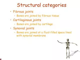
Classification of joints Joints SynarthrosisDiarthrosis FibrousCartilagenousSynovial
Synarthrosis A synarthrosis is a type of joint which permits very little or no movement under normal conditions. The synarthrosis occurs between the bones that is almost direct contact, fastened together by intervening connective tissue or hyaline cartilage, and in which there is no appreciable motion. It includes the Fibrous joints Cartilaginous joints Synostosis.
Fibrous Joints (Fixed joint) • The joints that are held together by dense fibrous tissue • No movement occurs in these joints except syndesmoses which allows little movement • Types of Fibrous Tissue: • Suture • Syndesmoses • Gomphosis
Sutures 1.Thin layer of dense fibrous connective tissue unites bones of the skull 2.Immovable (synarthroses) 3.If fuse completely in adults is synostosis
Syndesmoses • Type of fibrous joint where the bones are united by interoseous ligament & the bones concerned are some distance apart • Such ligaments persists throughout life and a slight movement is possible • e.g. Radioulnar joint, Tibiofibular joint.
Gomphosis • It is a specialized articulation restricted to the fixation of teeth in the maxilla & mandible
Cartilaginous Joints (slightly movable joint) • The joints that are held together by cartilage • Here a little amount of movement is possible. • 2 types • synchondrosis • symphysis
Synchondrosis 1. The Hyaline cartilage in these joint is temporary in nature & is replaced completely by bone. e.g. Between Epiphysis & Diaphysis of a growing long bone 2. Immovable 4. It is a temporary form of joint
Symphysis • Fibrocartilage is connecting material • 2. Slightly movable • 3. Intervertebral discs and pubic symphysis
Synostosis The bones are united by the ossification of the fibrous joint or synchondrosis between them, e. g. , the synosteosis between the ilium, ischium and pubis of the hip bones.
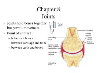
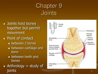
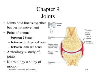
DiarthrosesSynovial Joints • Synovial jointsare freely movable joints that contain synovial fluid in a cavity surroundingthe ends of articulating bones.
Essential structures of synovial joints • Articular surface: covered by articular cartilage articular head articular fossa • Articular capsule • Fibrous membrane • Synovial membrane • Articular cavity: containing a trace of synovial fluid; subatmospheric pressure in it
Characteristics • The participating bones are held together by an articular capsule • It is composed of an outer fibrous capsule & an inner synovial membrane • Synovial membrane lines the whole of the interior of the joint cavity except the articular surface • Articular surface are covered by articular cartilage
Characteristics • They are separated by narrow space called joint cavity • Cavity contains a colourless, transparent, viscous fluid, rich in hyaluronic acid called synovial fluid • Joint cavity may contain some intra articular structures like articular disc, meniscus etc. • Movement is permitted from limited to a wide range.
Synovial membrane • Synovial membrane • inner lining of capsule • secretes synovial fluid containing hyaluronic acid (slippery) • brings nutrients to articular cartilage
Synovial Bursa • It is a closed connective tissue sac lined with synovial membrane filled with synovial fluid. Types • Subtendinous e.g. Biceps and subscapularis bursa • Articular e.g. subacromial bursa • Subcutaneous – e.g. pre patellar bursa Functions • Diminishes friction • Allows free movement • Helps in lubrication
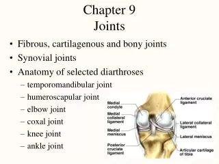
Intra articular Structures Cartilaginous structure • Articular Disc • Articular Menisci • GlenoidLabrum Ligaments traversing the joints • LigamentumTeres of hip joint • Cruciate ligament of Knee joint Muscle Tendon • Long head of Biceps- Shoulder Joint • Tendon of Popliteus- Knee Joint
Articular Disc / Articular Menisci • These are the fibro cartilaginous structures between articular surfaces dividing the joint cavity completely or incompletely • Articular Meniscus divides the joint incompletely into two compartments • Complete - e g. Sternoclavicular joint • Incomplete – e.g. Knee joint
Functions • Acts as a buffer & shock absorber • Strengthen the joint • Smooth in the articulation between the bony surfaces
Articular Labra • It is a fibrocartilaginous annular lip which is attached to the margin of an articular surface • E.g. Glenoid cavity & acetabulum
Articular fat pad • These are accumulation of adipose tissue present in many synovial joints • They are covered by synovial membrane E.g. Hip Joint • Makes the joint cavity uniform • Increase surface area of synovial joint
Types of synovial joints • Uniaxial joints: • hinge joints • trochoid (pivot) joints • Biaxial joints: • ellipsoid joints • saddle joints • Multiaxial joints: • ball-and-socket joint • plane joints
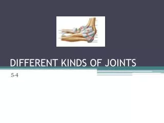
Uniaxial joints --Hinge Joint • Convex surface of one bones fits into concave surface of 2nd bone • Uniaxial like a door hinge • Examples • Knee, elbow, ankle, interphalangeal joints • Movements produced • flexion = decreasing the joint angle • extension = increasing the angle • hyperextension = opening the joint beyond the anatomical position
Uniaxial joints --Pivot Joint • Rounded surface of bone articulates with ring formed by 2nd bone & ligament • Monoaxial since it allows only rotation around longitudinal axis • Examples • Proximal radioulnar joint • supination • pronation • Atlanto-axial joint • turning head side to side “no”
Biaxial joints –Condyloid or Ellipsoidal Joint • Oval-shaped projection fits into oval depression • Biaxial = flex/extend or abduct/adduct is possible • Examples • wrist and metacarpophalangeal joints for digits 2 to 5
Adduction and Abduction Condyloid joints Ball and Socket joints
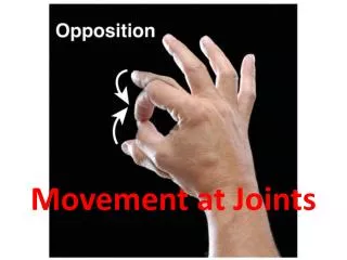
Biaxial joints – Saddle Joint • One bone saddled-shaped; other bone fits as a person would sitting in that saddle • Biaxial • Circumduction allows tip of thumb travel in circle • Opposition allows tip of thumb to touch tip of other fingers • Example • trapezium of carpus and metacarpal of the thumb
Multiaxial joints –Ball and Socket Joint • Ball fitting into a cuplike depression • Multiaxial • flexion/extension • abduction/adduction • rotation • circumduction • Examples • shoulder joint • hip joint
Multiaxial joints – Plane Joint • Bone surfaces are flat or slightly curved • Side to side movement only • Rotation prevented by ligaments • Examples • intercarpal or intertarsal joints • sternoclavicular joint • vertebrocostal joints
ⅠThe vertebral column 1 The vertebral joints nucleus pulposus annulus fibrosus The intervertebral disc The anterior longitudinal ligament The posterior longitudinal ligament The joints of the vertebral bodies Interspinal ligament Supraspinal ligament Intertransverse ligament The joint of articular process The joints of the vertebral arches
2 The vertebral column as a whole and its movements Anterior aspect Dorsal aspect Lateral aspect (4 curves) The vertebral column as a whole flexion and extension rotation circumduction The movement of the vertebral column
ⅡThe Thorax The costovertebral joints The costotransverse joints 1 Costovertebral joint 2 Sternocostal joints : true, false and floating rib Superior inferior 2 openings 3 walls infrasternal angle intercostal space 3 The thorax ( thoracic cage) as a whole
Articulations of Bones of Trunk The vertebral columnconsists of 24 vertebrae, the sacrum, and the coccyx.
Joints of the vertebral bodies Intervertebral discs between bodies of adjacent vertebrae, composed of: • Nucleus pulposus, an inner soft, pulpy, highly elastic structure (gelatinous core ) • Annulus fibrosus an outer fibrous ring consisting of fibrocartilage
Anterior longitudinal ligament • Strong band covering the anterior part of the vertebral bodies and intervertebral discs running from the anterior margin of foramen magnum to the S1~S2 • Maintains stability of the intervertebral disc and prevents hyperextension of the vertebral column Posterior longitudinal ligament • Attached to the posterior aspect of the intervertebral discs and posterior edges of the vertebral bodies from C2 vertebra to sacrum • Prevents hyperflexion of the vertebral column and posterior protrusion of the discs
Joints of the vertebral arches • Ligamentaflava― elastic ligament, unite laminae of adjacent vertebrae, and complete the posterior wall of vertebral canal; tend to prevent hyperflexion of the vertebral column • Interspinal ligament • Supraspinal ligament • Intertansverseligament
Atlantooccipital joint • Between superior articulating surfaces of atlas and occipital condyles • Supported by membrances and ligaments that join occipital bone and atlas • Action ― nodding of head, lateral tilting of head
Atlantoaxial joint • Three synovial joints between atlas and axis • Laterally, paired joints between articulating facets • Median joint between dens of axis and anterior arch of atlas • Supported by ligaments • apical ligament of dens • alar ligament • transverse ligament of atlas • tectorial membrane • Action ― allow atlas (and head) to pivot on the axis and vertebral column
Normal Curves of vertebral column • Cervical curvatureconvex forward • Thoracic curvatureconvex backward • Lumbar curvature convex forward • Sacral curvatureconvex backward Movement of the vertebral column • flexion • extension • lateral flexion • rotation
Thoracic cage Composition Bones ― consists of twelve thoracic vertebrae, twelve pairs of ribs and costal cartilages, and sternum
Joints • Costovertebraljoints • Joints of costal head • Costotransverse joints • Sternocostal joints • Sternocostalsynchondrosis of first rib • Sternocostaljoints • Interchondraljoints:between costal cartilages 8, 9, and 10 to form the costal arch
General features of thoracic cage • Roughly cone-shape, narrow above and broad below, flattened from before-backwards, longer behind than in front • Inlet of thorax: bounded by upper border of manubrium, first rib, and vertebra T1 • Outlet of thorax: bounded by vertebra T12, 12th and 11th ribs, costal arch and xiphoid process • Infrasternalangle: formed by the costal arch of both side • Intercostalspaces: lie between the ribs
Function: • protects the organs in the thoracic cavity and upper abdominal cavity; • plays a vital role in the process of breathing Expiration Inspiration
Joints of skull • Continuous joints: sutures, synchondrosis or synosteosis