Bleeding Disorders
Bleeding Disorders. 1. Thrombocytopenia Platelet (thrombocyte) deficiency Even normal movements can cause bleeding from small blood vessels that require platelets for clotting Petechiae – purple blotches 2. Hemophilia Hereditary bleeding disorder Normal clotting factors are missing


Bleeding Disorders
E N D
Presentation Transcript
Bleeding Disorders 1.Thrombocytopenia • Platelet (thrombocyte) deficiency • Even normal movements can cause bleeding from small blood vessels that require platelets for clotting • Petechiae – purple blotches 2. Hemophilia • Hereditary bleeding disorder • Normal clotting factors are missing • Sex-linked genetic disorder
3. Anemia a. Hemorrhagic – massive bleeding b. Hemolytic – bacterial infection c. Pernicious – lack of B12 absorption d. Aplastic – damage of bone marrow e. Iron-deficiency – diet, menstral flow, bleeding ulcer f. Sickle-cell – recessive genetic disorder; sickling due to increase O2 levels Bleeding Disorders
4. Polycythemia – abnormal increase of RBCs; from bone cancer or response to high altitude; causes high viscosity. 5. pH disorders – regulated by kidneys & resp. sys. a. alkalosis – too basic b. acidosis – too acidic Bleeding Disorders
Blood Groups and Transfusions • Large losses of blood have serious consequences • Loss of 15 to 30 percent causes weakness • Loss of over 30 percent causes shock, which can be fatal • Transfusions are the only way to replace blood quickly • Transfused blood must be of the same blood group
Human Blood Groups • Blood contains genetically determined proteins • A foreign protein (antigen) may be attacked by the immune system • Blood is “typed” by using antibodies that will cause blood with certain proteins to clump (agglutination)
Human Blood Groups • There are over 30 common red blood cell antigens • The most vigorous transfusion reactions are caused by ABO and Rh blood group antigens • If not matched blood types: Lysed RBCs release hemoglobin which blocks kidneys, causes fevers, vomit, etc.
ABO Blood Groups • Based on the presence or absence of two antigens (A & B) • Blood Types • Type A – has A antigen, anti-B antibody • Type B – has B antigen, anti-A antibody • Type AB – has both A & B antigens, Universal Recipient • Type O – lacks A & B antigens, has both anti-A anti-B antibodies; Universal Donor
Rh Blood Groups • Named because one of eight Rh antigens (agglutinogen D) found first in Rhesus monkey • Most Americans are Rh+ • Anti-Rh antibodies not automatically formed; formed after exposed to Rh+ • Problems can occur in mixing Rh+ blood into a body with Rh– blood
Rh Dangers During Pregnancy • Danger when Mom is Rh– and Dad is Rh+, and child inherits the Rh+ • Rh– mother carrying Rh+ baby can cause problems for the unborn child • The first pregnancy usually without problems • In a second pregnancy, the mother’s immune system produces antibodies to attack the Rh+ blood (hemolytic disease of the newborn) • Destruction of RBCs, anemia, brain damage, death • Fetal transfusions
Blood Typing • Blood samples are mixed with anti-A and anti-B serum • Coagulation or no coagulation leads to determining blood type • Typing for ABO and Rh factors is done in the same manner • Cross matching – testing for agglutination of donor RBCs by the recipient’s serum, and vice versa
Developmental Aspects of Blood • Sites of blood cell formation • The fetal liver and spleen are early sites of blood cell formation • Bone marrow takes over hematopoiesis by the 7th month • Fetal hemoglobin can pick up more O2 • Physiological Jaundice – liver can’t keep up with rapid fetal RBC destruction
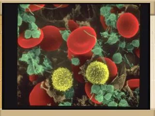
several nuclear lobes connected by thin filamentous strands of chromatin. Neutrophils
nucleus that often appears horseshoe or kidney shaped. The chromatin appears lacy and nucleoli are usually not apparent. The nucleus looks a bit like a "brain." Monocytes
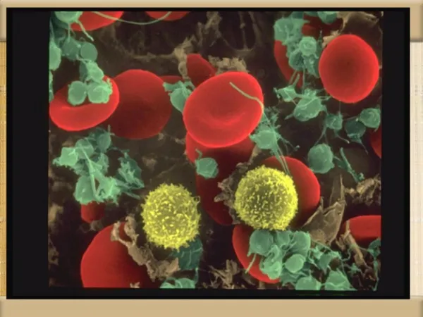
The nucleus rounded or oval, the same size as RBC. Chromatin densely packed with no apparent nucleoli. Lymphocyte nucleus almost always appears smudged. The cytoplasm is scanty and stains pale blue. Lymphocytes
nucleus consists of 2 to 3 lobes but is usually not as lobulated as neutrophils. The cytoplasm is full of dark purple specific granules Basophils
bilobate (two lobes) nucleus and a cytoplasm full of brightly stained eosinophilic (orange-red) specific granules Eosinophils & Platelets