Download
1 / 63
660 likes | 771 Views
Protein Aggregation and Disease. Examples of disorders that are involve protein accumulation in the ER: Congenital Hypothyroidism thyroglobulin accumulates (goiter) Liver damage associated with anti-trypsin accumulates in liver cells emphysema Osteogenesis Imperfecta type I pro-collagen

E N D
Protein Aggregation and Disease Examples of disorders that are involve protein accumulation in the ER: Congenital Hypothyroidism thyroglobulin accumulates (goiter) Liver damage associated with anti-trypsin accumulates in liver cells emphysema Osteogenesis Imperfecta type I pro-collagen Hereditary Hypofibrinogemia fibrinogen
Conformational Diseases Alzheimer’s Disease:Beta amyloid peptide Prion Diseases: Prion proteins (PrP) Parkinson’s Disease:a-synuclein aggregates to form major component of intracellular “Lewy Bodies” Amyotrophic lateral sclerosis: intracellular superoxide dismutase Huntington’s Disease:Huntingtin protein that contain long stretches of polyglutamine rare vs. inherited
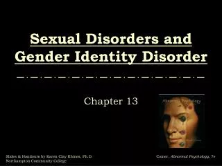
Hungtington’s Disease: Involves a Cytosolic Protein
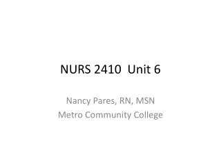
Alzheimer’s Disease
Alzheimer’s Disease Neuronal cell death triggered by aggregates of amyloid-b peptide. Long form (Ab42) especially aggregation-prone. In simple forms of AD, Ab42 overproduction occurs. In sporadic AD, “factors” promote amyloid-b aggregation by… increasing amount of amyloid-b decreasing degradation of amyloid-b decreasing transport of the amyloid-b out of the brain increasing Ab42:Ab40 ratio altering properties of amyloid-b
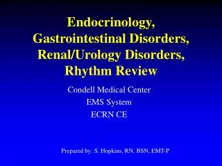
Possible Routes for Amyloid Formation and Cellular Toxicity Unfolded protein Folding intermediate Properly folded protein Vulnerable beta sheet structure aggregation factors that could promote amyloid formation at any step: aging, stress, local environment, cell type, toxins (e.g., Al(III), etc. “cross beta” fibrils cellular toxicity through any of a variety of possible mechanisms super-aggregates form and other molecules associate resulting in mature plaques
Protein-Ligand Interactions: Affinity and Specificity “Affinity” is a description of the tightness of binding. “High affinity” = tight binding = large Keq = large |-Go| = (usually) long lifetime for the complex. Proteins are capable of very high binding specificity.
Protein-Ligand Interactions The Simplest Case: 1:1 Stoichiometry R + L RL R: “receptor” L: “ligand” Equilibrium Constant: [RL] Kassociation = Ka = [R] . [L] [R] . [L] Kdissociation = Kd = [RL] Ka = 1/Kd DGoassociation = -DGodissociation
Kd or Ka? Kd is usually used because of its convenient units: mM, mM, or nM
“Dose/Response”Measurable output (response) will vary with L concentration (dose). D/R maximum when all R is complexed with L… “saturation” % of maximal possible functional response of R population is directly proportional to: fraction of R sites complexed (fR) = [RL] [L] = [R]total Kd + [L] When [L] >> Kd, then fR approaches 1… leading to maximum response.
This “binding isotherm” equation can be plotted: “saturation” 1.0 half maximal saturation 0.5 free ligand concentration required to achieve fR = 0.5 is equal to Kd 0 0 Maximum % change in fR per unit [L] is in the 0-0.5 range
Important implications and considerations… • It is free [L] which is being plotted. • However, if [L]free is approximately = [L]total, • then [L]total may be plotted. • To approach 100% saturation [L] must be >>Kd. • When [L] is<< 1-2 Kd, fR changes steeply. • This equation is a lot like the M-M equation: • fR v/Vmax • Kd Km • Dose-response and the [L] required to give 50%-maximum effect: • Ki, LD50, I50 or Kapparent
For a 150 lb. patient and … … a 300 g/mol drug that binds to a single protein target. Milligrams of Drug to Achieve 5 X Kd Kd 1 nM 0.1 1 μM 100 1 mM 100,000
Fitting a Model to Data If binding is involves 1:1 model, then data should be fit by a hyperbola. 1.0 0 0
If model is not appropriate, then the model will not be well fit to the data. 1.0 0 0
Typical Ranges for Dissociation ConstantsGo(kcal/mol) Enzyme-Substrate Interactions 1 mM to 5 mM -3 to -8 Receptor-Hormone Interactions 0.01 nM to 10 mM -7 to -15 Antibody-Antigen Interactions 0.01 nM to 10 mM -7 to -15 Dissociation constants often reflect the physiological concentrations of the ligands involved. Changes in amino acid sequence in a protein can tune affinity up or down.
intercept = 1/Kd slope = -1/Kd fR/[L] 0 0 1.0 fR Making Binding Data Linear [RL] [L] fR = = [R]total Kd + [L] Rearrange to: fR/[L] = 1/Kd – fR/Kd Scatchard Equation
Binding affinity and kinetics R + L RL Association rate = kon. [R] . [L] Dissociation rate = koff. [RL] at equilibrium: kon. [R] . [L] = koff. [RL] koff [R] . [L] = = Kd kon [RL] kon usually 105-108 M-.sec- koff usually scales directly with Kd: small Kd means low koff half-life of complex = t1/2 = 0.69/koff Affinity of 1 nM means t1/2 in the range of 10 sec to 2 hours. “Irreversible” Binding…
Myoglobin, Hemoglobin, and Oxygen: • Paradigms for Simple and Complex Binding • Physiologically important • Historically important • However… • “Partial pressure” units are unusual • Hemoglobin: • unusually complex • 5 mM concentration (30% w/v)
Hemoglobin Myoglobin
Myoglobin and Hemoglobin • Myoglobin (Mb) • monomeric globular protein • 1:1 binding stoichiometry • main function is to carry oxygen from capillaries to tissues • Hemoglobin (Hb) • Tetramer: two alpha and two beta subunits • alpha and beta subunits are homologous • alpha/beta structure similar • binds up to 4 molecules of O2 • binding is cooperative • binding also regulated by heteroallostery • function is to transport oxygen from lungs to capillaries, • also helps carry CO2 back to lungs • Both have porphryin cofactor. • Porphyrin contains an iron Fe(II) ion: complex called “heme”
Binding of oxygen to myoglobin. PO2: partial pressure of oxygen q : same as fR P50: same as Kd
Why Myoglobin Does Not Serve as the Primary Oxygen Delivery Vehicle of Blood PO2 in the lungs is 100 torr. PO2 in capillaries is ca. 30 torr.
Why Myoglobin Does Not Serve as the Primary Oxygen Delivery Vehicle of Blood PO2 in the lungs is 100 torr. PO2 in capillaries is ca. 30 torr.
Binding that is more complicated than 1:1 receptor:ligand stoichiometry. • The variables… • How many sites for a given ligand? • Are sites identical? • Are sites cooperative? • Is there a second type of ligand that also binds to the receptor? • Does it compete for the same site(s)? • Is there allostery? 1 ligand, 2 sites, homocooperative: R + L RL RLL K1 K2 K1[L] + 2K1K2[L]2 fR = 2 . (1 + K1[L] + K1K2[L]2)
If Ka,2 > Ka,1: “positive cooperativity” Homocooperative 2:1 L:R binding is the simplest route to a sigmoidal binding.
Hemoglobin has 4 binding sites, which are positively homocooperative. The Hill equation: [L]a fR = Kd,apparent + [L]a fR log = a. log[L] - logKd,apparent (1 – fR)
What is the physiological significance of cooperativity in hemoglobin?
Other Allosteric Effectors of Hemoglobin The O2 Binding/Delivery Properties of Hb Are Further Optimized By: H+: Bohr Effect(Negative Allostery) CO2 Release of CO2 into capillaries from respiring tissues lowers pH via conversion to HCO3- to reinfoce Bohr Effect CO2 + H2O HCO3- + H+ CO2 also chemically reacts with Hb in capillaries (Negative Allostery) Bisphosphoglycerate (BPG). Binding to Hb: Negative Allostery
Additive Allosteric Effects are Used To Achieve Optimal Binding/Delivery The Bohr Effect Applies in lungs. Applies in Capillaries
Hemoglobin with bound bisphosphoglycerate at b-b subunit interface.
b a b O2 a H+,CO2,BPG R (high affinity) T (low affinity) The Monod-Wyman-Changeux Model for Cooperativity in Hemoglobin
Effect of oxygen on heme iron(Major Trigger for Shift in the T to R State Equilibrium)
The T to R Cooperative Transition in Hemoglobin Blue = T state (deoxy) Red = R-state (oxy) and in the b/a interface
Deoxyhemoglobin (T State) Oxyhemoglobin (R State)
Low affinity for O2 High affinity for O2 Regulation of Hemoglobin TR Keq Keq : Depends on concentrations of… O2: shifts Keq to the right H+, CO2, and BPG: shifts Keq to the left Lungs: High O2, Low CO2 Capillaries: Lower O2, Lower pH, High CO2 BPG: Promotes O2 dissociation in caps much more than it inhibits O2 binding in lungs.
Purposes for Allostery/Cooperativity in Proteins • Control of Loading/Delivery • On/Off Switch • Throttle • Metabolic Feedback
Relationship of Hemoglobin Genetics and Human Disease • ψ : pseudogenes (not transcribed) • ζ,ε : embryonic Hb chains • : fetal Hb subunit (fetal Hb = 22) Adult Hb: 22(96%), 2δ2 (3%), 22 (1%) note that there are four alleles for the chain, but only a pair for
Hemoglobin-Based Disorders • Thalessemias: Are caused by the failure to produce one or more Hb • chains in a functional form. • β-Thalessemia major: failure to produce • the Hb-β chain. Fatal. • β-Thalessemia minor :ca. 50% of the usual • amount of the Hb-β. Mild. • Hemoglobin Variants: • Hundreds Known • Most from missense mutations • Most are “neutral mutations ” (a.k.a. “neutral polymorphisms”) • Sometimes result in disease… such as sickle cell anemia.
Sickle Cell Anemia • First documented “molecular disease”. • An inherited disorder. • Hemoglobin S: Glu6Val mutation in beta subunit. • Hemoglobin S: it is the deoxy form that is prone to polymerize. • A recessive disorder. (Mutations must be homozygous). • Heterozygous case confers resistance to malaria. • 30% of sub-Saharan Africans and 8% of African-Americans • carry HbS allele… also common in Latin-Americans • No cure yet, but there are therapies (e.g., hydroxyurea). • Today’s “Clinical Correlation”…