Epithelial Tissue
Epithelial Tissue. Key Terms. Histology: the study of tissues. Tissues: Simply groups of similar cells that work together performing the same task Greatest form of teamwork in the body. Where are tissues found?. With few exceptions, organs are composed of four basic tissue types:

Epithelial Tissue
E N D
Presentation Transcript
Key Terms • Histology: • the study of tissues. • Tissues: • Simply groups of similar cells that work together performing the same task • Greatest form of teamwork in the body
Where are tissues found? With few exceptions, organs are composed of four basic tissue types: – Epithelial Tissue – Connective Tissue – Muscular Tissue – Nervous Tissue
Why Study Histology? Knowing the difference between normal and abnormal tissue is the first step in diagnosis and treatment of patients.
Skin, our largest organ * made of all four tissue types
Epithelial Tissue • Makes up 3% of your body weight • They don’t move • They don’t send messages • Their cells are all touching one another • Of all tissues, they are the most widely varied in structure and function
Locations of Epithelial Tissues • Covers the body (epidermis) • Found on the inside of hollow organs and the outside of allorgans • Found above a connective tissue layer (epi = above) • Lines the cavities, tubes, ducts, and blood vessels inside the body
Epithelial Anatomy • Apical surface – upper surface that is free or exposed to the “exterior” • Basal surface – attached surface (below) • Microvilli – small fingerlike extensions that increase the surface area allowing for more work to be done
Functions of Epithelial Tissue • Protects from physical & chemical injury • Protects against microbial infection • Contains nerve endings which respond to stimuli • Filters, secretes & reabsorbs materials • Secretes fluids to lubricate joints
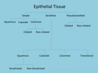
Three Basic Shapes • Squamous – like scales, or pancakes (“being squashed like a pancake”) • Cuboidal – looks like cubes • Columnar – longer and look like columns
Cell Organization • Simple – single layer of cells; typically found where absorption and filtration occur or a single layer of epithelial is needed simple squamous simple cuboidal simple columnar • Stratified – layers of cells; common in areas where protection is needed like the skin stratified squamous stratified cuboidal stratified columnar
Two Types of Stratified Columnar • Ciliated • Unciliated cilia No cilia
Squamous Epithelium • Simple – one cell thick • Forms solid layer of cells which line blood vessels, body cavities and covers organs in body cavities • Stratified – multiple layers • Forms epidermis
Duct Duct Cuboidal Epithelium Cuboid Cells • Simple – one cell thick • Roughly cube shaped • Line ducts in kidneys where re-absorption and secretion activities take place. Cuboid Cells
Columnar Epithelium • Simple – one cell thick • Column shaped (long and narrow) • Lines digestive tract where re-absorption & secretion occurs.
Confusing Epithelial Tissue • Transitional Epithelium – stratified tissue that can’t make up its mind as to whether it is squamous or cuboidal • Shape of cells depends upon the amount of stretching (ex: bladder)
Confusing Epithelial Tissue Continued… • “Pseudostratified Columnar Epithelium” • Looks like it has more than one layer because of the position of the nucleus • Nuclei are positioned at differing levels • Cells narrow in the area without the nucleus
Types of Epithelial Membranes • Mucous or mucosa– lining of tubes; moistens and protects from enzymes (stomach, trachea, and vagina) • Serous or serosa – outside of organs; lubricates (all thoracic, abdominal and pelvic organs) • Cutaneous or skin – body surface; protection • Synovial – synovial joints; lines and protects synovial cavities (elbow, knee, hip, etc.)
Synovial Membranes • Some joints are surrounded by a membrane (synovium) that produces a thick, synovial fluid. This fluid nourish the cartilage and keeps it slippery. Knee Joint
Mucosa Membranes • Mucus-secreting membrane that lines all body cavities or passages that come in contact with the outside of the body. Membranes are involved in absorption and secretion. Fungus
Serous Membranes • Serous membranes line body cavities that do not open directly to the outside, and they cover the organs located in those cavities. Fluid lubricates the membrane and reduces friction and abrasion when organs move against each other. Canine Reproductive Organs
Cutaneous Membranes • Cutaneous membranes of the skin cover the surface of the body. They consist of stratified squamous epithelium and the underlying connective tissues. Cutaneous membranes are thick, relatively waterproof, and dry. Skin Cancer