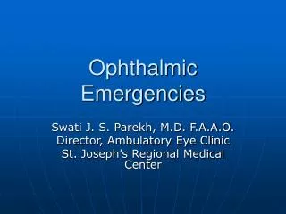
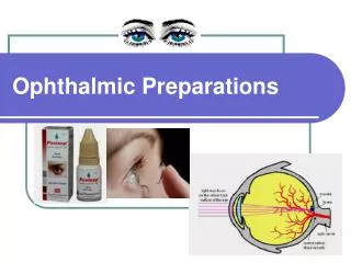
Ophthalmic Preparations
Ophthalmic Preparations. Ophthalmic preparations:. Ophthalmic preparation Applied topically to the cornea, or instilled in the space between the eyeball and lower eyelid Solution Dilute with tear and wash away through lacrimal apparatus Administer at frequent intervals Suspension

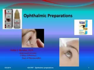
Ophthalmic Preparations
E N D
Presentation Transcript
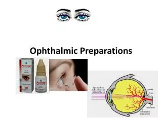
Ophthalmic preparations: • Ophthalmic preparation • Applied topically to the cornea, or instilled in the space between the eyeball and lower eyelid • Solution • Dilute with tear and wash away through lacrimal apparatus • Administer at frequent intervals • Suspension • Longer contact time • Irritation potential due to the particle size of drug • Ointment • Longer contact time and greater storage stability • Producing film over the eye and blurring vision
Ophthalmic preparations: • Controlled delivery system • Release at a constant rate for a long time • Enhanced corneal absorption • Drug with not serious side effect or tolerate by the patient
Drugs used in the eye: • Miotics e.g. pilocarpine Hcl • Mydriatics e.g. atropine • Cycloplegics e.g. atropine • Anti-inflammatories e.g. corticosteroids • Anti-infectives (antibiotics, antivirals and antibacterials) • Anti-glucoma drugs e.g. pilocarpine Hcl • Surgical adjuncts e.g. irrigating solutions • Diagnostic drugs e.g. sodiumfluorescein • Anesthetics e.g. tetracaine
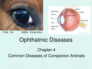
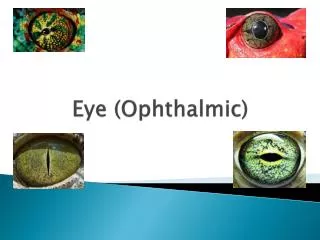
Anatomy and Physiology of the Eye: • Human eye • Diameter of 23 mm • Three layers: • Outermost coat : the clear, transparent cornea and the white, opaque sclera • Middle layer : the iris anteriorly, the choroid posteriorly, and the ciliary body at the intermediate part • Inner layer : retina (extension of CNS)
Anatomy and Physiology of the Eye (Cont.): • The sclera: The protective outer layer of the eye, referred to as the “white of the eye” and it maintains the shape of the eye. • The cornea: The front portion of the sclera, is transparent and allows light to enter the eye. The cornea is a powerful refracting surface, providing much of the eye's focusing power. • The choroid is the second layer of the eye and lies between the sclera and the retina. It contains the blood vessels that provide nourishment to the outer layers of the retina. • The iris is the part of the eye that gives it color. It consists of muscular tissue that responds to surrounding light, making the pupil, or circular opening in the center of the iris, larger or smaller depending on the brightness of the light.
Anatomy and Physiology of the Eye (Cont.): • The lens is a transparent, biconvex structure, encased in a thin transparent covering. The function of the lens is to refract and focus incoming light onto the retina. • The retina is the innermost layer in the eye. It converts images into electrical impulses that are sent along the optic nerve to the brain where the images are interpreted. • The macula is located in the back of the eye, in the center of the retina. This area contains the highest concentration of cones, produces the sharpest vision.
Anatomy and Physiology of the Eye (Cont.): • The inside of the eyeball is divided by the lens into two fluid-filled sections. • The larger section at the back of the eye is filled with a colorless gelatinous mass called the vitreous humor. • The smaller section in the front contains a clear, water-like material called aqueous humor. • The conjunctiva is a mucous membrane that begins at the edge of the cornea and lines the inside surface of the eyelids and sclera, which serves to lubricate the eye.
Absorption of drugs in the eye: Factors affecting drug availability: 1- Rapid solution drainage by gravity, induced lachrymation, blinking reflex, and normal tear turnover: • The normal volume of tears = 7 ul, the blinking eye can accommodate a volume of up to 30 ul without spillage, the drop volume = 50 ul
Absorption of drugs in the eye: 2- Superficial absorption of drug into the conjunctiva and sclera and rapid removal by the peripheral blood flow: 3- Low corneal permeability In general: • Transport of hydrophilic and macromolecular drugs through scleral route • Lipophilic agents of low molecular weight follow transcorneal transport by passive diffusion and obey Ficks‘s first law of diffusion: J = - D . dCm / dx
Corneal absorption: J = The flux rate across the membrane D = diffusion coefficient - The diffusion coeffecient , as the molecular size of the drug Cm = concentration gradient • As the drug solubility , the gradient , the driving force for drug entry into the aqueous humor N.B.the drug should have dual solubility (oil and water soluble) to traverse the corneal epithelium (lipid barrier) then the aqueous humour.
General safety considerations: A. Sterility: • Ideally, all ophthalmic products would be terminally sterilized in the final packaging. - Only a few ophthalmic drugs formulated in simple aqueous vehicles are stable to normal autoclaving temperatures and times (121°C for 20-30 min). *Such heat-resistant drugs may be packaged in glass or other heat-deformation-resistant packaging and thus can be sterilized in this manner. • Most ophthalmic products, however cannot be heat sterilized due to the active principle or polymers used to increase viscosity are not stable to heat.
A. Sterility (cont.): * Most ophthalmic products are aseptically manufactured and filled into previously sterilized containers in aseptic environments using aseptic filling-and-capping techniques.
B. Ocular toxicity and irritation: • Albino rabbits are used to test the ocular toxicity and irritation of ophthalmic formulations. • The procedure based on the examination of the conjunctiva, the cornea or the iris. • E.g. USP procedure for plastic containers: 1- Containers are cleaned and sterilized as in the final packaged product. 2- Extracted by submersion in saline and cottonseed oil. 3- Topical ocular instillation of the extracts and blanks in rabbits is completed and ocular changes examined.
C.Preservation and preservatives: • Preservatives are included in multiple-dose eye solutions for maintaining the product sterility during use. • Preservatives not included in unit-dose package. • The use of preservatives is prohibited in ophthalmic products that are used at the of eye surgery because, if sufficient concentration of the preservative is contacted with the corneal endothelium, the cells can become damaged causing clouding of the cornea and possible loss of vision. so these products should be packaged in sterile, unit-of-use containers. • The most common organism is Pseudomonas aeruginosa that grow in the cornea and cause loss of vision.
C.Preservation and preservatives: Examples of preservatives: 1- Cationic wetting agents:• Benzalkonium chloride (0.01%) • It is generally used in combination with 0.01-0.1% disodium edetate (EDTA). The chelating, EDTA has the ability to render the resistant strains of PS aeruginosa more sensitive to benzalkonium chloride. 2- Organic mercurials:• Phenylmercuric nitrate 0.002-0.004% phenylmercuric acetate 0.005-0.02%.
C.Preservation and preservatives: 3-Esters of p-hydroxybenzoic acid:• Mixture of 0.1% of both methyl and propyl hydroxybenzoate (2 :1) 4- Alcohol Substitutes:• Chlorobutanol(0.5%). Effective only at pH 5-6.• Phenylethanol (0.5%)
Manufacturing considerations: A. Manufacturing Environment: The environment should be sterile and particle-free through: -Laminar-flow should be used throughout the manufacturing area. -Total particles per cubic foot of space should be minimum. - Relative humidity controlled to between 40 and 60%. - Walls, ceilings and floors should be constructed of materials that are hard, non flaking, smooth and non-affected by surface cleaners or disinfectants.
A. Manufacturing Environment: • Ultraviolet lamps provided in flush-mounted fixtures to maintain surface disinfection • Separate entrance for personnel and equipment should be provided through specially designed air locks that are maintained at negative pressure relative to the aseptic manufacturing area and at a positive pressure relative to the noncontrolled area
B. Manufacturing Techniques: • Aqueous ophthalmic solutions:
B. Manufacturing Techniques: • Aqueous suspensions:
B. Manufacturing Techniques: • Ophthalmic ointment:
B. Manufacturing Techniques: • Unpreserved formulations of active drug(s):
Ideal ophthalmic delivery system: Following characteristics are required to optimize ocular drug delivery system: • Good corneal penetration. • Prolong contact time with corneal tissue. • Simplicity of instillation for the patient. • Non irritative and comfortable form • Appropriate rheological properties
CLASSIFICATION OF OCULAR DRUG DELIVERY SYSTEMS: • Ointments • Gels • Topical eye drops: • Solutions • - Suspensions • - Powders for • reconstitution • - Sol to gel systems - Ocular inserts
A. Topical Eye drops: • Administration: • Pull down the eyelid • Tilting the head backwards • Look at the ceiling after the tip is pointed close to the lower cul-de-sac • Apply a slight pressure to the rubber bulb or plastic bottle to allow a drop to fall into the eye. To prevent contamination: • Clean hands • Do not touch the dropper tip to the eye and surrounding tissue • Do not squeeze lids
A. Topical Eye drops: 1- Solutions: - Ophthalmic solutions are sterile solutions, essentially free from foreign particles, suitably compounded and packaged for instillation into the eye.
Disadvantages of the eye drops: 1-The very short time the solution stays at the eye surface. The retention of a solution in the eye is influenced by viscosity, hydrogen ion concentration, the osmolality and the instilled volume. 2- its poor bioavailability (a major portion i.e. 75% is lost via nasolacrimal drainage) 3- the instability of the dissolved drug 4- the necessity of using preservatives.
2- suspensions: to prevent irritation or scratching of the Cornea.
Isotonicity Lacrimal fluid is isotonic with blood having an isotonicity value Corresponding to that of 0.9% Nacl solution
2- pH Adjustment and Buffers: • pH adjustment is very important as pH affects: 1- to render the formulation more stable 2- The comfort, safety and activity of the product. Eye irritation increase in tear fluid secretion Rapid loss of medication. 3- to enhance aqueous solubility of the drug. 4- to enhance the drug bioavailability 5- to maximize preservative efficacy