Chromosomes
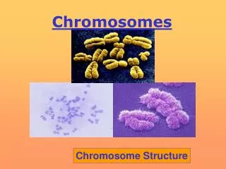
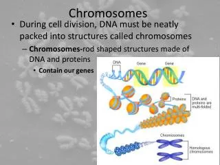
Chromosomes. Dr. R. Siva VIT University, INDIA rsiva77in@rediffmail.com. What Exactly is a chromosome?. Chromosomes are the rod-shaped , filamentous bodies present in the nucleus , which become visible during cell division . They are the carriers of the gene or unit of heredity.

Chromosomes
E N D
Presentation Transcript
Chromosomes Dr. R. Siva VIT University, INDIA rsiva77in@rediffmail.com
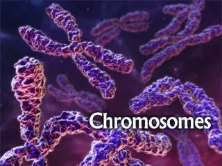
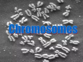
What Exactly is a chromosome? Chromosomes are the rod-shaped, filamentous bodies present in the nucleus, which become visible during cell division. They are the carriers of the gene or unit of heredity. Chromosome are not visible in active nucleus due to their high water content, but are clearly seen during cell division.
Chromosomes were first described by Strausberger in 1875. • The term “Chromosome”, however was first used by Waldeyer in 1888. • They were given the name chromosome (Chromo = colour; Soma = body) due to their marked affinity for basic dyes. • Their number can be counted easily only during mitotic metaphase.
Chromosomes are composed of thin chromatin threads called Chromatin fibers. • These fibers undergo folding, coiling and supercoiling during prophase so that the chromosomes become progressively thicker and smaller. • Therefore, chromosomes become readily observable under light microscope. • At the end of cell division, on the other hand, the fibers uncoil and extend as fine chromatin threads, which are not visible at light microscope
Number of chromosomes • Normally, all the individuals of a species have the same number of chromosomes. • Closely related species usually have similar chromosome numbers. • Presence of a whole sets of chromosomes is called euploidy. • It includes haploids, diploids, triploids, tetraploids etc. • Gametes normally contain only one set of chromosome – this number is called Haploid • Somatic cells usually contain two sets of chromosome - 2n : Diploid
3n – triploid 4n – tetraploid The condition in which the chromosomes sets are present in a multiples of “n” is Polyploidy When a change in the chromosome number does not involve entire sets of chromosomes, but only a few of the chromosomes - is Aneuploidy. • Monosomics (2n-1) • Trisomics (2n+1) • Nullisomics (2n-2) • Tetrasomics (2n+2)
Organism No. chromosomes • Human 46 • Chimpanzee 48 • Dog 78 • Horse 64 • Chicken 78 • Goldfish 94 • Fruit fly 8 • Mosquito 6 • Nematode 11(m), 12(f) • Horsetail 216 • Sequoia 22 • Round worm 2
Organism No. chromosomes • Onion 16 • Mold 16 • Carrot 20 • Tomato 24 • Tobacco 48 • Rice 24 • Maize 20 • Haploppus gracilis 4 • Crepis capillaris 6
On the extreme, round worm shows only two chromosomes, while the other extreme is represented by Protozoa having 300 or more chromosomes. • However, most organisms have numbers between 12 to 50. • 3-8 in fungi • From 8 – 16 in Angiosperms (Most common number being 12).
Chromosome Size • In contrast to other cell organelles, the size of chromosomes shows a remarkable variation depending upon the stages of cell division. • Interphase:chromosome are longest & thinnest • Prophase:there is a progressive decrease in their length accompanied with an increase in thickness • Anaphase: chromosomes are smallest. • Metaphase: Chromosomes are the most easily observed and studied during metaphase when they are very thick, quite short and well spread in the cell. • Therefore, chromosomes measurements are generally taken during mitotic metaphase.
The size of the chromosomes in mitotic phase of animal and plants sp generally varies between 0.5 µ and 32 µ in length, and between 0.2 µ and 3.0 µ in diameter. The longest metaphase chromosomes found in Trillium- 32 µ. The giant chromosomes found in diptera and they may be as long as 300 µ and up to 10 µ in diameter. In general, plants have longer chromosomes than animal and species having lower chromosome numbers have long chromosomes than those having higher chromosome numbers Among plants, dicots in general, have a higher number of chromosome than monocots. Chromosomes are longer in monocot than dicots.
In order to understand chromosomes and their function, we need to be able to discriminate among different chromosomes. • First, chromosomes differ greatly in size • Between organisms the size difference can be over 100-fold, while within a sp, some chromosomes are often 10 times as large as others. • In a species Karyotype, a pictorial or photographic representation of all the different chromosomes in a cell of an individual, chromosomes are usually ordered by size and numbered from largest to smallest.
Can distinguish chromosomes by “painting” – using DNA hybridization + fluorescent probes – during mitosis
Karyotype: is the general morphology of the somatic chromosome. Generally, karyotypes represent by arranging in the descending order of size keeping their centromeres in a straight line. • Idiotype: the karyotype of a species may be represented diagrammatically, showing all the morphological features of the chromosome; such a diagram is known as Idiotype.
Chromosomes may differ in the position of the Centromere, the place on the chromosome where spindle fibers are attached during cell division. • In general, if the centromere is near the middle, the chromosome is metacentric • If the centromere is toward one end, the chromosome is acrocentric or submetacentric • If the centromere is very near the end, the chromosome is telocentric.
The centromere divides the chromosome into two arms, so that, for example, an acrocentric chromosome has one short and one long arm, • While, a metacentric chromosome has arms of equal length. • All house mouse chromosomes are telocentric, while human chromosomes include both metacentric and acrocentric, but no telocentric.
Autosomal pair Sex chromosome DiploidNo. of No. of X Y (2n) metacentrics acrocentric or telocentric Cat 38 16 2 M M Dog 78 0 38 M A Pig 38 12 6 M M Goat 60 0 29 A M Sheep 54 3 23 A M Cow 60 0 29 M M Horse 64 13 18 M A M – Metacentric; A – Acrocentric
Euchromatin and Heterochromatin • Chromosomes may be identified by regions that stain in a particular manner when treated with various chemicals. • Several different chemical techniques are used to identify certain chromosomal regions by staining then so that they form chromosomal bands. • For example, darker bands are generally found near the centromeres or on the ends (telomeres) of the chromosome, while other regions do not stain as strongly. • The position of the dark-staining are heterochromatic region or heterochromatin. • Light staining are euchromatic region or euchromatin.
Heterochromatin is classified into two groups: (i) Constitutive and (ii) Facultative. • Constitutive heterochromatin remains permanently in the heterochromatic stage, i.e., it does not revert to the euchromatic stage. • In contrast, facultative heterochromatin consists of euchromatin that takes on the staining and compactness characteristics of heterochromatin during some phase of development.
Satellite DNAs When the DNA of a prokaryote, such as E.coli, is isolated, fragmented and centrifuged to equilibrium in a Cesium chloride (CsCl) density gradient, the DNA usually forms a single band in the gradient. On the other hand, CsCl density-gradient analysis of DNA from eukaryotes usually reveals the presence of one large band of DNA (usually called “Mainband” DNA) and one to several small bands. These small bands are referred to as “Satellite DNAs”. For e.g., in Drosophila virilis, contain three distinct satellite DNAs.
Not only the genomes of eukaryotes are more complex than prokaryotes, but the DNA of eukaryotic cell is also organized differently from that of prokaryotic cells. • The genomes of prokaryotes are contained in single chromosomes, which are usually circular DNA molecules. • In contrast, the genomes of eukaryotes are composed of multiple chromosomes, each containing a linear molecular of DNA.
Although the numbers and sizes of chromosomes vary considerably between different species, their basic structure is the same in all eukaryotes Organism Genome Chromosome Size (Mb)a numbera Arabidopsis thaliana 70 5 Corn 5000 10 Onion 15,000 8 Lily 50,000 12 Fruit fly 165 4 Chicken 50,000 39 Mouse 1,200 20 Cow 3000 30 Human 3000 23 a – both genome size and chromosome numbers are for haploid cells
The DNA of eukaryotic cell is tightly bound to small basic proteins (histones) that package the DNA in an orderly way in the cell nucleus. • This task is substantial (necessary), given the DNA content of most eukaryotes • For e.g., the total extended length of DNA in a human cell is nearly 2 m, but this must be fit into a nucleus with a diameter of only 5 to 10µm. • Although DNA packaging is also a problem in bacteria, the mechanism by which prokaryotic DNA are packaged in the cell appears distinct from that eukaryotes and is not well understood.
Prokaryotic chromosome • The prokaryotes usually have only one chromosome, and it bears little morphological resemblance to eukaryotic chromosomes. • Among prokaryotes there is considerable variation in genome length bearing genes. • The genome length is smallest in RNA viruses • In this case, the organism is provided with only a few genes in its chromosome. • The number of gene may be as high as 150 in some larger bacteriophage genome.
In E.coli, about 3000 to 4000 genes are organized into its one circular chromosome. • The chromosome exists as a highly folded and coiled structure dispersed throughout the cell. • The folded nature of chromosome is due to the incorporation of RNA with DNA. • There are about 50 loops in the chromosome of E.coli. • These loops are highly twisted or supercoiled structure with about four million nucleotide pairs. • Its molecular weight is about 2.8 X10⁹ • During replication of DNA, the coiling must be relaxed. • DNA gyrase is necessary for the unwinding the coils.
Bacterial Chromosome • Single, circular DNA molecule located in the nucleoid region of cell
Supercoiling Most common type of supercoiling The helix twists on itself; twists to the right Helix twists on itself in the opposite direction; twists to the left
Mechanism of folding of a bacterial chromosome There are many supercoiled loops (~100 in E. coli) attached to a central core. Each loop can be independently relaxed or condensed. Topoisomerase enzyme – (Type I and II) that introduce or remove supercoiling.
Chromatin • The complexes between eukaryotic DNA and proteins are called Chromatin, which typically contains about twice as much protein as DNA. • The major proteins of chromatin are the histones – small proteins containing a high proportion of basic aminoacids (arginine and lysine) that facilitate binding negatively charged DNA molecule . • There are 5 major types of histones: H1, H2A, H2B, H3, and H4 – which are very similar among different sp of eukaryotes. • The histones are extremely abundant proteins in eukaryotic cells. • Their mass is approximately equal to that of the cell’s DNA
The major histone proteins: Histone Mol. Wt No. of Percentage Amino acid Lys + Arg H1 22,500 244 30.8 H2A 13,960 129 20.2 H2B 13,774 125 22.4 H3 15,273 135 22.9 H4 11,236 102 24.5 The DNA double helix is bound to proteins called histones. The histones have positively charged (basic) amino acids to bind the negatively charged (acidic) DNA. Here is an SDS gel of histone proteins, separated by size
In addition, chromatin contains an approximately equal mass of a wide variety of non-histone chromosomal proteins. • There are more than a thousand different types of these proteins, which are involved in a range of activities, including DNA replication and gene expression. • The DNA of prokaryotes is similarly associated with proteins, some of which presumably function as histones do, packing the DNA within the bacterial cell. • Histones, however are unique feature of eukaryotic cells and are responsible for distinct structural organization of eukaryotic chromatin
The basic structural unit of chromatin, the nucleosome, was described by Roger Kornberg in 1974. • Two types of experiments led to Kornberg’s proposal of the nucleosome model. • First, partial digestion of chromatin with micrococcal nuclease (an enzyme that degrades DNA) was found to yield DNA fragments approximately 200 base pairs long. • In contrast, a similar digestion of naked DNA (not associated with protein) yielded a continuous smear randomly sized fragments. • These results suggest that the binding of proteins to DNA in chromatin protects the regions of DNA from nuclease digestion, so that enzyme can attack DNA only at sites separated by approximately 200 base pairs.
Electron microscopy revealed that chromatin fibers have a beaded appearance, with the beads spaced at intervals of approximately 200 base pairs. • Thus, both nuclease digestion and the electron microscopic studies suggest that chromatin is composed of repeating 200 base pair unit, which were called nucleosome.
Detailed analysis of these nucleosome core particles has shown that they contain 146 base pairs of DNA wrapped 1.75 times around a histone core consisting of two molecules each of H2A, H2B, H3, and H4 (the core histones). • One molecule of the fifth histone H1, is bound to the DNA as it enters and exists each nucleosome core particle. • This forms a chromatin subunit known as chromatosome, which consist of 166 base pairs of DNA wrapped around histone core and held in place by H1 (a linker histone)
Centromeres and Telomeres • Centromeres and telomeres are two essential features of all eukaryotic chromosomes. • Each provide a unique function i.e., absolutely necessary for the stability of the chromosome. • Centromeres are required for the segregation of the centromere during meiosis and mitosis. • Teleomeres provide terminal stability to the chromosome and ensure its survival
Centromere • The region where two sister chromatids of a chromosome appear to be joined or “held together” during mitatic metaphase is called Centromere • When chromosomes are stained they typically show a dark-stained region that is the centromere. • Also termed as Primary constriction • During mitosis, the centromere that is shared by the sister chromatids must divide so that the chromatids can migrate to opposite poles of the cell. • On the other hand, during the first meiotic division the centromere of sister chromatids must remain intact • whereas during meiosis II they must act as they do during mitosis. • Therefore the centromere is an important component of chromosome structure and segregation.
As a result, centromeres are the first parts of chromosomes to be seen moving towards the opposite poles during anaphase. • The remaining regions of chromosomes lag behind and appear as if they were being pulled by the centromere.
Kinetochore • Within the centromere region, most species have several locations where spindle fibers attach, and these sites consist of DNA as well as protein. • The actual location where the attachment occurs is called the kinetochore and is composed of both DNA and protein. • The DNA sequence within these regions is called CEN DNA.
Typically CEN DNA is about 120 base pairs long and consists of several sub-domains, CDE-I, CDE-IIandCDE-III. • Mutations in the first two sub-domains have no effect upon segregation, • but a point mutation in the CDE-III sub-domain completely eliminates the ability of the centromere to function during chromosome segregation. • Therefore CDE-III must be actively involved in the binding of the spindle fibers to the centromere.
The protein component of the kinetochore is only now being characterized. • A complex of three proteins called Cbf-IIIbinds to normal CDE-III regions but can not bind to a CDE-III region with a point mutation that prevents mitotic segregation.