Download
1 / 4
40 likes | 170 Views
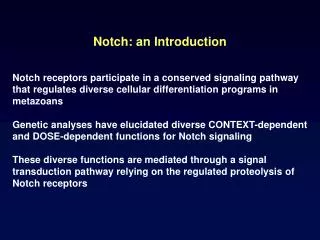
Tb 3+. Zn 2+. Ca 2+. Domain Organization of the Notch Receptors and the Notch Signaling Pathway. Crystal Structure of LNR and HD Domain of human Notch2 1. NMR Structure of hN1LNRA 2. N15. Figure B. Figure C. C4. C9. C22. Ca 2+. C27. D30. D33. N-term. S19. C34. C18. C-term.

E N D
Tb3+ Zn2+ Ca2+ Domain Organization of the Notch Receptors and the Notch Signaling Pathway Crystal Structure of LNR and HD Domain of human Notch21 NMR Structure of hN1LNRA2 N15 Figure B Figure C C4 C9 C22 Ca2+ C27 D30 D33 N-term S19 C34 C18 C-term Figure A Characterization of the Ca2+ Binding Affinity and Coordination Site of the LIN-12/Notch-Repeat Christina Hao, Didem Vardar-Ulu Wellesley College Chemistry Department Wellesley, Massachusetts • Introduction • Notch receptors are transmembrane glycoproteins that regulate cell fate in multicellular organisms via a highly conserved signaling pathway (Figure A). • Deregulation of notch signaling pathway in all four identified notch homologues (Notch1 – Notch4) has been implicated in numerous disease phenotypes. • Three conserved Lin12/Notch Repeat (LNRA, LNRB, and LNRC) modules are located in tandem in the extracellular region of the notch receptors. They maintain the receptor in a resting conformation prior to ligand-induced activation. (Figure B). • Each LNR module in the Notch receptor consists of three characteristic disulfide bonds and a Ca2+ ion, essential for structural integrity (Figure C). Results Representations of overall structures and calcium binding sites for (A) hN1LNRA (B) Glucose Transferase LNRA (C) hN4LNRA (A)NMR Structure. (B) and (C) homology Modeling Ca2+ dependency during folding of hN1 LNRA, glucose transferase and hN4LNRA B. Glucose Transferase LNRA C. hN4 LNRA A. hN1 LNRA • Key: • Calcium binding sites (7 angstroms from the Ca2+): red and green ribbons • Red sticks: Aspartates - Green sticks: other residues besides aspartates - Blue sphere: calcium ion • Non-binding sites: silver ribbons Representative ITC data on the calorimetric titrations of hN1LNRA with Ca2+,Zn2+ ,Tb3+ • Objectives • Quantify calcium binding affinity and specificity of LNR homologues across different proteins using ITC • Determine calcium dependency of different LNR homologues for autonomous folding • Understand the molecular basis of calcium binding in LNR using computer modeling Distances and distribution of coordinating residues from Ca2+ Sequence Alignment of LNR homologues investigated in this study: Key: Red residues: residues that coordinate calcium with both side chain and backbone oxygen moiety Distances ranges highlighted yellow: aspartate is present in this distance range • Conclusions • HN1 LNRA exclusively binds to Ca2+ in an exothermic reaction with a dissociation constant of 22.05 +/- 3.27 µM and a stoichiometry of 1:1 at pH 7.0. • Glucose transferase LNRA display strong binding to calcium in a non-stoichiometric manner • HN4 LNRA does not bind to calcium • Calcium is necessary for the folding of HN1 LNRA and glucose transferase LNRA but not for HN4 LNRA • Homology modeling suggests differences in distribution of aspartic acids lead to distinct calcium binding behaviors of the LNR repeats. Representative ITC data on the calorimetric titrations of glucose transferase and hN4LNRA with Ca2+ • Material and Methods • Protein Acquisition: • Human N1LNRA recombinantly expressed in Escherichia coli. Human Notch 4 LNRA and glucose transferase LNRA were synthesized by EZ Biolabs. • All proteins were folded and purified as follows: • Folding: 6-8 hours dialysis against refolding buffer: 2.5mM cysteine, 0.5mM cystine, 100mM NaCl, 200mM sucrose, 10mM CaCl2 and 20mM Tris pH 8. • Purification: Elution through reverse phase high pressure liquid chromatography (RP-HPLC) using 0.1% formic acid in acetonitrile based buffer systems. Peaks corresponding to the folded species were collected and lyophilized. • Folding Experiments: • Denatured proteins were refolded in redox solution containing 5:1 cysteine/cystine ratio, 100mM NaCl and 20mM Tris at pH 8 and with/without 10mM CaCl2. Proteins were folded under partial nitrogen atmosphere for six hours and promptly analyzed on RP-HPLC. • Isothermal titration calorimetry (ITC) Experiments: • Lyophilized protein of appropriate concentration was demetalized with sigma chelex beads and suspended in 35mM Hepes pH7, 100mM NaCl buffer . • Stock metal solution of 0.2 – 1mM CaCl2 was used • Isothermal titration calorimetry experiments (ITC), were carried out using a high-precision VP-ITC titration calorimetry instrument (Microcal Inc., Northampton, MA) where the metal solution was titrated in 5µL increments into the protein solution at 20°C. • Computer Modeling Software used: • Clustal W: sequence alignment • Modeller: homology modeling • Pymol: visualization Glucose transferase LNRA hN4 LNR A hN1 LNR A • Future Directions • Determine precise roles of disulphide bonds and aspartic acids in calcium binding affinity through mutational studies. • Correlate calcium binding affinity and specificity with structural stability to gain insight into the biological significance of calcium binding by the LNRs in vivo. • Design of calcium binding peptides through de novo experiments based on understanding of the LNRs Summary of thermodynamic parameters associated with the binding of Ca2+ to Zn2+ and Tb3+ presaturated hN1LNRA • References • Gordon, W. R.;* Vardar-Ulu, D.;* Histen, G.; Sanchez-Irizarry, C.; Aster, J. C.; Blacklow, S. C. “Structural basis for autoinhibition of Notch” Nat Struct Mol Biol.2007, 14, 295–300.2. • Vardar, D.; North, C. L.; Sanchez-Irizarry, C.; Aster, J. C.; Blacklow, S. C. “NMR Structure of a Prototype LNR Module from Human Notch1” Biochemistry2003, 42, 7061–7067. • N. Eswar, M.A. Marti-Renom, b. Webb, m.S. Madhusudhan, D. Eramian, M. Shen, U. Pieper, A. Sali, Comparative Protein Structure Modeling with MODELLER. Current Protocols in Bioinformatics, John Wilery & Sons, Inc., Supplement 15, 5.6. 1-5.6.30, 2000 • Cheng G, Baker D and Samudrala R. A Novel Small Molecule Crystal Structure Derived Potential Function To Predict Protein Metal Ion Binding Site, Affinity and Specificity From Structure. xxxx.YYYY,aa-bb,2007 • http://protinfo.compbio.washington.edu/soak/