Download
1 / 10
100 likes | 377 Views
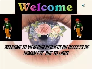
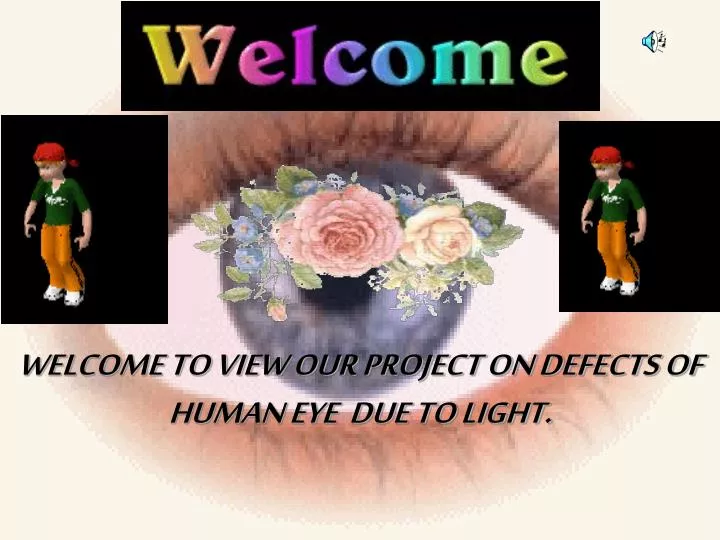
WELCOME TO VIEW OUR PROJECT ON DEFECTS OF HUMAN EYE DUE TO LIGHT. INTRODUCTION

E N D
WELCOME TO VIEW OUR PROJECT ON DEFECTS OF HUMAN EYE DUE TO LIGHT.
INTRODUCTION Eye, light-sensitive organ of vision in animals. The eyes of various species vary from simple structures that are capable only of differentiating between light and dark to complex organs—such as those of humans and other mammals—that can distinguish minute variations of shape, color, brightness, and distance. The actual process of seeing is performed by the brain rather than by the eye. The function of the eye is to translate the electromagnetic vibrations of light into patterns of nerve impulses that are transmitted to the brain.
DEFECTS OF THE EYE Myopia:(nearsightedness) This is a defect of vision in which far the refractive power of the eye’s lens too strong. objects appear blurred but near objects are seen clearly. The image is focused in front of the retina rather than on it usually because the eyeball is too long or Myopia can be corrected by wearing glasses/contacts with concave lenses these help to focus the image on the retina Hyperopia:(farsightedness) This is a defect of vision in which there is difficulty with near vision but far objects can be seen easily. The image is focused behind the retina rather than upon it. This occurs when the eyeball is too short or the refractive power of the lens is too weak. Hyperopia can be corrected by wearing glasses/contacts that contain convex lenses.
Astigmatism: This defect is when the light rays do not all come to a single focal point on the retina, instead some focus on the retina and some focus in front of or behind it. This is usually caused by a non-uniform curvature of the cornea. A typical symptom of astigmatism is if you are looking at a pattern of lines placed at various angles and the lines running in one direction appear sharp whilst those in other directions appear blurred. Astigmatism can usually be corrected by using a special spherical cylindrical lens; this is placed in the out-of-focus axis.
DIFFERENT FOCUS OF EYE Focusing the Eye Light rays entering the eye are refracted, or bent, when they pass through the lens. Normal vision requires that the rays focus on the retina. If the eyeball is too long, an accurately focused image falls short of the retina. This is called myopia, or shortsightedness. A shortsighted person sees distant objects unclearly. Longsighted focus, or hyperopia, results when the eyeball is too short. In this case, an accurately focused image would fall behind the retina.
CONCAVE LENS A concave lens is curved inward; it is shaped like two dishes placed back-to-back. Light passing through a concave lens bends outward, or diverges. Unlike convex lenses, which produce real images, concave lenses produce only virtual images. A virtual image is one from which light rays only appear to come. This one appears as a smaller image just in front of the actual object (in this case a shamrock). Concave lenses are generally prescribed for myopic, or short-sighted, people. Concave lenses help the eyes to produce a sharp image on the retina instead of in front of it.
CONVEX LENS A convex lens has a thick centre and thinner edges. Light passing through a convex lens is bent inward, or made to converge. This causes an image of the object to form on a screen on the opposite side of the lens. The image is in focus if the screen is placed at a particular distance from the lens that depends upon the distance of the object and the focal point of the lens. The lens in the human eye is convex, but, unlike a glass lens, it is elastic so that it can change shape to focus on objects at varying distances. The lens becomes short and fat when viewing close objects and elongated and thin when viewing distant objects.
SCANNING ELECTRON MICROSCOPE This scanning electron microscope (SEM) is to the left of the operator, with the computer images of the specimen on the screens to the right. Although the SEM cannot ”see“ objects as small as those that a transmission electron microscope can resolve, the images it produces are more useful for seeing the three-dimensional surface structure of small objects.
MAGNIFYING GLASS . A magnifying glass is a large convex lens commonly used to examine small objects. The lens bends incoming light virtual so that an enlarged, image ofthe object (in this case a mushroom) appears beyond it. The image is called virtual because the light rays that appear to come from it do not actually pass through it.