Development of IRMSI-MED: Advancing Synchrotron Infrared Microspectroscopy for Biological Imaging
The IRMSI-MED facility has been developed to enable rapid chemical imaging of biological single cells in vivo, providing critical insights into sub-cellular structure functions and responses to environmental changes. With its novel design featuring multiple detection channels, IRMSI-MED enhances the ability to discern fine chemical compositions and monitor dynamic shifts within cells over minutes. This versatile tool is available for a range of scientific disciplines including biology, nanoscience, and environmental science, fostering interdisciplinary research and education.

Development of IRMSI-MED: Advancing Synchrotron Infrared Microspectroscopy for Biological Imaging
E N D
Presentation Transcript
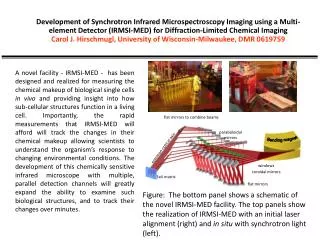
Development of Synchrotron Infrared Microspectroscopy Imaging using a Multi-element Detector (IRMSI-MED) for Diffraction-Limited Chemical Imaging Carol J. Hirschmugl, University of Wisconsin-Milwaukee, DMR 0619759 A novel facility - IRMSI-MED - has been designed and realized for measuring the chemical makeup of biological single cells in vivo and providing insight into how sub-cellular structures function in a living cell. Importantly, the rapid measurements that IRMSI-MED will afford will track the changes in their chemical makeup allowing scientists to understand the organism’s response to changing environmental conditions. The development of this chemically sensitive infrared microscope with multiple, parallel detection channels will greatly expand the ability to examine such biological structures, and to track their changes over minutes. flat mirrors to combine beams paraboloidalmirrors Bending magnet common optical axis Figure: The bottom panel shows a schematic of the novel IRMSI-MED facility. The top panels show the realization of IRMSI-MED with an initial laser alignment (right) and in situ with synchrotron light (left). windows toroidal mirrors 3x4 matrix flat mirrors
Development of Synchrotron Infrared Microspectroscopy Imaging using a Multi-element Detector (IRMSI-MED) for Diffraction-Limited Chemical Imaging Carol J. Hirschmugl, University of Wisconsin-Milwaukee, DMR 0619759 • During the realization of IRMSI-MED, Dr. El Bayarri (Jordan), who is directing the assembly of an IR facility at SESAME (Synchrotron in the Middle East to promote Peace and Science), participated in the assembly of the IRMSI-MED facility. • High School Teachers from Milwaukee Public Schools participating in RET at UWM have arranged visits for their students to SRC where they will use IRMSI-MED for demonstration experiments. • IRMSI-MED microscope will be available for users across a wide array of disciplines (e.g. soft matter condensed physics, nanoscience, biology, chemistry, veterinary science, engineering, environmental science and geology), providing a new interdisciplinary tool to the broader scientific community. Dr. El Bayarri (SESAME, Jordan) working on alignment issues during assembly of IRMSI-MED. “At Risk” High School Students visiting the SRC and running experiments at the IR Facility.