
TRPV5 Gene Organization and Constructs: Promoter Study in Mouse Kidney Cells
This study delves into the genomic organization of the TRPV5 gene with emphasis on the TRPV5 promoter region, including constructs like pAcGFP1-N1 plasmid, elucidated through detailed flow charts and immunolabeling in various kidney segments such as DCT, CNT, and CCD. The examination extends to the development of DCT2/CNT-specific AQP2 knockout mice. This comprehensive analysis sheds light on the intricate regulation and expression of relevant genes in renal tubules.

TRPV5 Gene Organization and Constructs: Promoter Study in Mouse Kidney Cells
E N D
Presentation Transcript
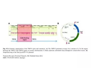
Fig. 15.1 Genomic organization of the TRPV5 gene and constructs. (a) The TRPV5 promoter (orange box) consists of a 3.6 kb region between the TRPV5 and TRPV6 genes on mouse chromosome 6. Peaks represent calculated Gerp intraspecies conservation scores. (b) Simplified map of the final pAcGFP1-N1 plasmid Transient Receptor Potential Channels, Md. Shahidul Islam (Ed.) ISBN: 978-94-007-0264-6, Springer
Fig. 15.2 Flow chart of the validation of the TRPV5 promoter driven EGFP expressing transgenic mouse line Transient Receptor Potential Channels, Md. Shahidul Islam (Ed.) ISBN: 978-94-007-0264-6, Springer
Fig. 15.3 TRPV5 promoter-driven EGFP expression in DCT2, CNT, and CCD. (a) Tripleimmunolabeling confocal microscopical analysis of fixed kidney sections for calbindin-D28 K, Na+-Cl– cotransporter (NCC), and EGFP. (b) Similar immuno-fluorescence labeling for aquaporin-2 (AQP2), NCC, and EGFP. (c) Immuno-fluorescence labeling for vacuolar type proton pump (H+-ATPase), AQP2, and EGFP. (d) Schematic representation of the renal tubular system with indication of the tubule segments and expression sites for EGFP, NCC, calbindin-D28 K, AQP2, and H-ATPase Transient Receptor Potential Channels, Md. Shahidul Islam (Ed.) ISBN: 978-94-007-0264-6, Springer
Fig. 15.4 (a) Map of the pBS185 plasmid containing the TRPV5 promoter fragments, including restriction sites. (b) Flow chart of the development of DCT2/CNT-specific AQP2 knockout mice Transient Receptor Potential Channels, Md. Shahidul Islam (Ed.) ISBN: 978-94-007-0264-6, Springer