Download
1 / 24
461 likes | 1.56k Views
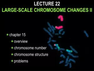
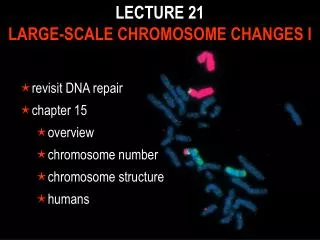
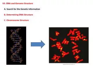
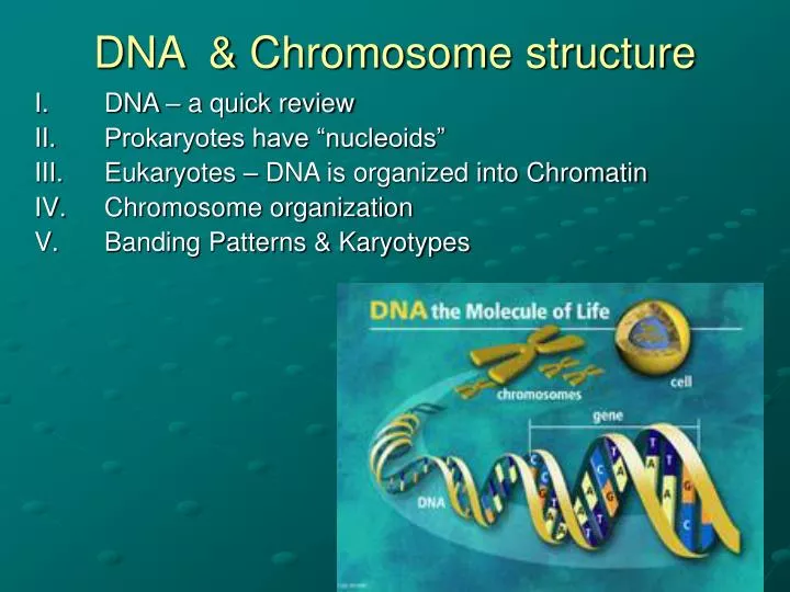
DNA & Chromosome structure. DNA – a quick review Prokaryotes have “nucleoids” Eukaryotes – DNA is organized into Chromatin Chromosome organization Banding Patterns & Karyotypes. I. DNA review. What are the criterion for genetic material? How does DNA structure = function?

E N D
DNA & Chromosome structure • DNA – a quick review • Prokaryotes have “nucleoids” • Eukaryotes – DNA is organized into Chromatin • Chromosome organization • Banding Patterns & Karyotypes
I. DNA review • What are the criterion for genetic material? • How does DNA structure = function? • How was DNA discovered to be the genetic material? • Why is a primer needed for replication? http://www.usfca.edu/fac-staff/dever/genetics_figure.htm
II. Chromosomes - Prokaryotes have “nucleoids” • Bacterial nucleoid = DNA + protein • DNA is in the form of loops, which are supercoiled and emerge from a dense protein containing structure:scaffold • DNA-binding proteins – structurally similar to histones (found in eukaryotes), have positively charged amino acids that bond to phosphate groups • HU • H
Supercoiled DNA, packed tighter…
III. Eukaryotes – DNA is organized into Chromatin • Eukaryotic DNA is highly organized by binding in a controlled manner to special proteins. • DNA in 1 chromosome of a human cell = 19,000 to 73,000 um long (as compared to 1,200 um in bacteria) • Histones & Nucleosomes are the key to packaging DNA into chromosomes. • Chromatin = nucleoprotein structure, loose • Chromsomes = only visible during mitosis, most packed state
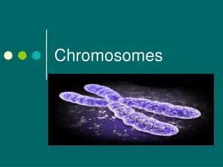
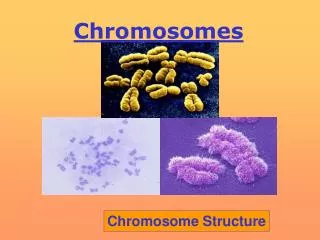
Chromosomes • Structures that contain the DNA for proper distribution of the genetic material during cell division
III. Chromosome organization there are several levels of organization – DNA 1.solenoid 2. chromatin fiber 3. scaffold 4. chromosome
1. Solenoid Hollow contact helix of nucleosomes (nucleosomes in one turn of the helix are in contact w/those of the next) 30nm fiber probably accounts for most of the chromatin in interphase.
2. chromatin fiber 3. scaffold 4. chromosome
Review: DNA is wrapped around spools called nucleosomes. Each nucleosome is composed of an octamer of proteins called histones. The DNA-nucleosome chain is further coiled and supercoiled into a 30nm structure known as a solenoid. The 30nm structure forms a series of looped domains that further condensed into the chromatin fiber 300 nm in diameter. The fibers are then coiled into the chromosome arms that constitute a chromatid.
A. Different types of chromatin: • Euchromatin: regions that stain more lightly because the genes are less compact • Heterochromatin: Dark stained regions, DNA is compact and thus genes are not transcribed
B. Chromatin Remodeling DNA – not static as chromatin, but moves between more condensed to less condensed state, for transcription purposes!
Review question • How is prokaryotic DNA packaging different from eukaryotic DNA packaging? Why so different? Why similar?
IV. Banding Patterns & Karyotypes Cytogenetics – field of genetics that involves the examination of chromosomes. • Banding: Certain chemical treatments of mammalian chromosomes yield differentially stained regions on chromosomes. The patterns obtained depend on the treatment used. • C-banding stains centromeres. • R-banding is the reverse of C-banding and stains non-centromeric regions in preference to centromeres. • G-banding is obtained with Giemsa stain. It yields a series of lightly and darkly stained bands • Q-banding is a fluorescent pattern obtained using quinacrine for staining. The pattern of bands is very similar to that seen in G-banding • Banding Differentiates regions along the chromosome: • characteristic series of lateral bands in each member of the chromosome set… • Used to identify homologous pairs of chromosomes
Banding – Chromosomes vary in size, centromere position and banding pattern! G banded Karyotype
B. Visible chromosome landmarks Centromere – constricted region of chromosome Telomere – end of chromosome
p arm – short arm of the chromosome; q arm – long arm of the chromosome • Arrangement of genes on chromosomes genes are the functional regions along the DNA molecule that constitutes a chromosome – the regions that are transcribed to produce RNA. • A chromosome represents large numbers of genes in a specific linear array & this array is different for different chromosomes. • Genes vary enormously in size • There are intergeneic segments • There are repeating segments
C. Chromosome Number: Different species have highly characteristic numbers of chromosomes. Chromosome number is the product of two other numbers: The haploid number and the number of sets. Diploid # or 2n = 46, Haploid # or n = 23
D. Sister chromatids & Homologous chromosomes • Homologues = members of a pair of chromosomes • for a diploid organism each type of chromosome is found in a homologous pair consisting of two parallel sister chromatids • If a particular gene is found on one chromosome; it is also found on the homologue