Download
1 / 1
10 likes | 238 Views
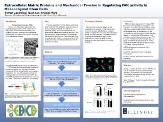
P=0.002264. P=0.958231. P=0.301495. P=0.721078. Fibronectin. Type I Collagen. Soft. Hard. Soft. Hard. Soft. Hard. Soft. Hard. 1h after seeding. 24h after seeding. Extracellular Matrix Proteins and Mechanical Tension in Regulating FAK activity in Mesenchymal Stem Cells.

E N D
P=0.002264 P=0.958231 P=0.301495 P=0.721078 Fibronectin Type I Collagen Soft Hard Soft Hard Soft Hard Soft Hard 1h after seeding 24h after seeding Extracellular Matrix Proteins and Mechanical Tension in Regulating FAK activity in Mesenchymal Stem Cells Type I Collagen Fibronectin 1 h 1 h 1 h 1 h Forrest Goodfellow, Taejin Kim, Yingxiao Wang Department of Bioengineering, College of Engineering, University of Illinois at Urbana-Champaign 0.6 kPa 40 kPa 40 kPa 0.6 kPa 24 h 24 h 24 h 24 h Introduction Focal Adhesion Kinase (FAK) FAK is a 125 kD protein responsible for tyrosine phosphorylation. FAK is activated in response to environmental stimuli such as extracellular matrix proteins, and mechanical tension. FAK can exits in they cytosol or bound to the plasma membrane. . Fluorescent Resonance Energy Transfer (FRET) FRET biosensors are genetically engineered proteins containing two fluorescent protein sequences without disrupting the functionality of the protein. The Lyn-FAK biosensor allows for the visualization of membrane bound FAK and Cyto-FAK allows for the visualization of cytosolic FAK. FRET biosensors enable live cell imaging. Aim Previous experiments in the Wang Laboratory have observed FAK activation under an array of environmental conditions. The effects of substrate rigidity and the composition of the extracellular matrix were observed with the Cyto-FAK biosensor. Now, the intent is to investigate FAK activation with the Lyn-FAK biosensor. This investigation will allow for the observation of FAK activity within the cytosol as well as bound to the plasma membrane. Preliminary Results The Ctyo-FAK biosensor allows for the visualization of cytosolic FAK activation and numerical analysis with Metaflour imaging software enables the numerical characterization of FAK activation with the Cyto-FAK biosensor. On going experiments will allow for the comparison of Cyto-FAK and Lyn-FAK biosensors under identical environmental conditions. Conclusions FAK activation observed with the Lyn-FAK biosensor demonstrated an increase in FAK activation on harder substrates relative to soft substrates, and the FAK activation at 1 hour differs significantly. An explanation for the difference may be the kinetics of FAK activation caused by the different ECM proteins. FAK activation must be observed with the Lyn-FAK biosensor before any further insights can be made. Continued research will allow for an understand of FAK distribution with in the cell and answer several questions. Is FAK synthesized in response to the environment? Does FAK transiently bind to the plasma membrane? Do ECM proteins effect the kinetics of FAK activation? 0.6 kPa 40 kPa 0.6 kPa 40 kPa a b www.spinal-research.org/ c Method www.neuroniccollagen.com/ www.nationaldiagnostics.com/ • Figure 1: (a) Polyacrylamide gel is used to simulate in virto environments of different mechanical tensions by varying the amount of cross-linked molecules (b) EM images of fibronectin and (c) collagen are ECM proteins responsible for cellular adhesion and play a role in FAK activation. Citations Seong J, Ouyang M, Kim T, Sun J, Wen PC, Lu S et al. Detection of focal adhesion kinase activation at membrane microdomains by fluorescence resonance energy transfer. Nat Commun 2011; 2: 406. Wang Y, Shyy JY, Chien S. Annu Rev Biomed Eng. 2008;10:1. Figure 3: FRET Images captured with Cyto-FAK biosensor. Panels represent Fibronectin and Type I Collagen coated gel substrate dishes at 1 and 24 hours after hMSC were plated on the gel. The stiffness of the substrate is indicated in the lower left corner. Acknowledgments EBICS Research Experience for Undergraduates Beckman Institute for Advance Science and Technology Jihye seong, et al. Figure 2: (a) indicates the design of the FAK biosensor and the phosphorylated tyrosine residue in red (b) Activated FAK will phosphorlate the biosensor’s substrate peptide and the proximity of ECPF and Ypet effects a change in FRET fluoresces. Figure 4: FRET ratios were calculated with Metaflour software. The FAK activation observed with the Cyto-FAK FRET ratio.