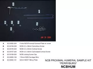
310 sample kit
Adenovirus IgA ELISA Kit can be provided from Creative Diagnostics.t<br>https://www.creative-diagnostics.com/Adenovirus-IgA-EIA-Kit-3835-498.htm<br>

310 sample kit
E N D
Presentation Transcript
SPECIFICATION SHEET Rev 110821EA-002CXY Adenovirus IgA ELISA Kit Prod. No.: DEIA310 Pkg. Size: 96T PRINCIPLE OF THE TEST INTENDED USE The Adenovirus IgA Antibody ELISA Test Kit has been de- signed for the the detection and the quantitative determina- tion of specific IgA antibodies against Adenovirus in serum and plasma. Further applications in other body fluids are pos- sible and can be requested from the Technical Service. This assay is intended for in-vitro diagnostic use only. Laboratory results can never be the only base of a medical report. The patient history and further tests have additionally to be taken into account. The Adenovirus IgA antibody test kit is based on the principle of the enzyme immunoassay (EIA). Adenovirus antigen is bound on the surface of the microtiter strips. Diluted patient serum or ready-to-use standards are pipetted into the wells of the microtiter plate. A binding between the IgA antibodies of the serum and the immobilized Adenovirus antigen takes place. After a one hour incubation at room temperature, the plate is rinsed with diluted wash solution, in order to remove unbound material. Then ready-to-use anti-human-IgA peroxi- dase conjugate is added and incubated for 30 minutes. After a further washing step, the substrate (TMB) solution is pipet- ted and incubated for 20 minutes, inducing the development of a blue dye in the wells. The color development is termi- nated by the addition of a stop solution, which changes the color from blue to yellow. The resulting dye is measured spectrophotometrically at the wavelength of 450 nm. The con- centration of the IgA antibodies is directly proportional to the intensity of the color. GENERAL DESCRIPTION The adenovirus is an ubiquitous pathogen of humans and animals. Adenoviruses are characterized by location inside the cell nucleus, common complement-fixing antigens and marked stability to environmental effects. Adenoviruses are endemic in all populations throughout the year. The infection is spread both through the aerial-droplet route and the routes characteristic for intestinal infections. The incubation period is between five and seven days. Adenoviruses mainly infest respiratory and intestinal mucosa, but also the cornea. They are accumulated in the epithelial cells and regional lymph nodes. Adenoviruses cause the widest variety of illnesses of the known respiratory viruses. The adenovirus infection is the most frequently caused viral disease of the respiratory tract among preschool children (types 1 - 5 and 7). Acute diseases of the upper respiratory tract occur predominantly. Pneumo- nia is the most severe form of adenoviral infection occurring mostly in infants below the age of one. Adenoviruses also cause outbreaks of swimmingpool- associated pharyngocon- junctival fever in the summer and epidemics of kerato- conjunctivitis of both children and adults. The intestinal form of adenoviral infection occurs mostly in children below the age of one. An acute adenoviral infection can be detected by virus isolation and/or serology. The serologic tests are par- ticularly important because they document actual infection in the patient and can be applied to large-scale epidemiologic investigations. The CF and ELISA tests measure predomi- nantly the antibodies directed against the group-specific de- terminants on the hexon component. The recommended tests for measuring type-specific antibodies are hemagglutinin inhi- bition and serum neutralization. The type-specific antigenic determinants of adenoviruses are located at the fibers on the capsid. Because of the ubiquity of the adenoviruses and nu- merous cross-reactions between related serotypes, serocon- version involving a fourfold or greater rise in antibody infec- tion is necessary to document infection. IgG is the predomi- nant antibody class measured in the serologic tests. REAGENTS PROVIDED Store kit components at 2-8°C and do not use after the expiry date on the box outer label. Before use, all components should be allowed to warm up to ambient temperature (18- 25°C). After use, the plate should be resealed, the bottle caps replaced and tightened and the kit stored at 2-8°C. The opened kit should be used within three months. Microtiter Strips: 12 strips with 8 breakable wells each, coated with a Adenovirus antigen (Adenovirus Hexon). Ready -to-use. Calibrator A (Negative Control): 2 mL, protein solution di- luted with PBS, contains no IgA antibodies against Adenovi- rus. Addition of 0.01 % methylisothiazolone and 0.01 % bro- monitrodioxane. Ready-to-use. Calibrator B (Cut-Off Standard): 2 mL human serum diluted with PBS, contains a low concentration of IgA antibodies against Adenovirus. Addition of 0.01 % methylisothiazolone and 0.01 % bromonitrodioxane. Ready-to-use. Calibrator C (Weak Positive Control): 2 mL, human serum diluted with PBS, contains a medium concentration of IgA antibodies against Adenovirus. Addition of 0.01 % methyli- sothiazolone and 0.01 % bromonitrodioxane. Ready-to-use. Calibrator D (Positive Control): 2 mL, human serum diluted with PBS, contains a high concentration of IgA antibodies against Adenovirus. Addition of 0.01 % methylisothiazolone and 0.01 % bromonitrodioxane. Ready-to-use. Enzyme Conjugate: 15 mL, anti-human-IgA-HRP (rabbit), in protein-containing buffer solution. Addition of 0.01 % methyli- sothiazolone and 0.01 % bromonitrodioxane and 5 mg/L Pro- clinTM. Ready-to-use. Creative Diagnostics. All rights reserved. 45-16 Ramsey Road Shirley, NY 11967, USA Tel: 631-624-4882 ·Fax:631-614-7828 E-mail: info@creative-diagnostics.com www.creative-diagnostics.com
Substrate: 15 mL, TMB (tetramethylbenzidine). Ready-to- use. Stop Solution: 15 mL, 0.5 M sulfuric acid. Ready-to-use. Sample Diluent: 60 mL, PBS/BSA buffer. Addition of 0.095 % sodium azide. Ready-to-use. Washing Buffer: 60 mL, PBS + Tween 20, 10x concentrate. Final concentration: dilute 1+9 with distilled water. If during the cold storage crystals precipitate, the concentrate should be warmed up at 37°C for 15 minutes. Plastic Foils: 2 pieces to cover the microtiter strips during the incubation. Plastic Bag: Resealable, for the dry storage of non-used strips. 2) Pipet 100 μL each of the diluted (1:101) samples and the ready-to-use standards and controls respectively into the wells. Leave one well empty for the substrate blank. 3) Cover plate with the enclosed foil and incubate at room temperature for 60 minutes. 4) Empty the wells of the plate (dump or aspirate) and add 300 μL of diluted washing solution. This procedure is re- peated totally three times. Rests of the washing buffer are afterwards removed by gentle tapping of the microtiter plate on a tissue cloth. 5) Pipet 100 μL each of ready-to-use conjugate into the wells. Leave one well empty for the substrate blank. 6) Cover plate with the enclosed foil and incubate at room temperature for 30 minutes. 7) Empty the wells of the plate (dump or aspirate) and add 300 μL of diluted washing solution. This procedure is re- peated totally three times. Rests of the washing buffer are afterwards removed by gentle tapping of the microtiter plate on a tissue cloth. 8) Pipet 100 μL each of the ready-to-use substrate into the wells. This time also the substrate blank is pipetted. 9) Cover plate with the enclosed foil and incubate at room temperature for 20 minutes in the dark (e.g. drawer). 10) To terminate the substrate reaction, pipet 100 μL each of the ready-to-use stop solution into the wells. Pipet also the substrate blank. 11) After thorough mixing and wiping the bottom of the plate, perform the reading of the absorption at 450 nm (optionally reference wavelength of 620 nm). The color is stable for at least 60 minutes. MATERIALS REQUIRED BUT NOT PROVIDED 1. 5 μL-, 100 μL- and 500 μL micro- and multichannel pipets 2. Microtiter Plate Reader (450 nm) 3. Microtiter Plate Washe 4. Reagent tubes for the serum dilution 5. Bidistilled water SPECIMEN COLLECTION AND HANDLING Principally serum or plasma (EDTA, citrate) can be used for the determination. Serum is separated from the blood, which is aseptically drawn by venipuncture, after clotting and cen- trifugation. The serum or plasma samples can be stored re- frigerated (2-8°C) for up to 48 hours, for a longer storage they should be kept at -20°C. The samples should not be frozen and thawed repeatedly. Lipemic, hemolytic or bacterially con- taminated samples can cause false positive or false negative results. For the performance of the test the samples (not the calibrators) have to be diluted 1:101 with ready-to-use sample diluent (e.g. 5 μL serum + 500 μL sample diluent). EVALUATION The mean values for the measured absorptions are calcu- lated after subtraction of the substrate blank value. The differ- ence between the single values should not exceed 10%. 1. Qualitative Evaluation The calculated absorptions for the patient sera, as mentioned above, are compared with the value for the cut-off standard. If the value of the sample is higher, there is a positive result. For a value below the cut-off standard, there is a negative result. It seems reasonable to define a range of +/-20 % around the value of the cut-off as a grey zone. In such a case the repetition of the test with the same serum or with a new sample of the same patient, taken after 2-4 weeks, is recom- mended. Both samples should be measured in parallel in the same run. The positive control must show at least the double absorption compared with the cut-off standard. 2. Quantitative Evaluation The ready-to-use standards and controls of the Adenovirus antibody kit are defined and expressed in arbitrary units (U/ mL). This results in an exact and reproducible quantitative evaluation. Consequently for a given patient follow-up con- trols become possible. The values for controls and standards in units are printed on the labels of the vials. For a quantita- tive evaluation the absorptions of the standards and controls are graphically drawn against their concentrations. From the ASSAY PROCEDURE 1. Reagent And Sample Preparation Washing Solution: dilute before use 1+9 with distilled water. If during the cold storage crystals precipitate, the concentrate should be warmed up at 37°C for 15 minutes. 1) Strict adherence to the protocol is advised for reliable per- formance. Any changes or modifications are the responsibility of the user. 2) All reagents and samples must be brought to room tem- perature before use, but should not be left at this temperature longer than necessary. 3) Standards and samples should be assayed in duplicates. 4) A standard curve should be established with each assay. 5) Return the unused microtiter strips to the plastic bag and store them dry at 2-8°C. 2. Assay Steps 1) Prepare a sufficient amount of microtiter wells for the stan- dards, controls and samples in duplicate as well as for a sub- strate blank. Creative Diagnostics. All rights reserved. 45-16 Ramsey Road Shirley, NY 11967, USA Tel: 631-624-4882·Fax:631-614-7828 E-mail: info@creative-diagnostics.com www.creative-diagnostics.com
resulting reference curve the concentration values for each patient sample can then be extracted in relation to their ab- sorptions. It is also possible to use automatic computer pro- grams. REFERENCES 1. Hierholzer JC, Tannock GA. Adenoviruses. In: Rose RR, Friedman H, Fahey JL. Manual of Clinical Laboratory Im- munology (third edition). American Society for Microbiol- ogy, Washington D.C. 527 (1986). 2. Mc Minn PC, Stewart J, Burrell CJ. A Community Out- break of Epidemic Keratoconjunctivitis in Central Australia Due to Adenovirus Type 8. J. Inf. Dis., 164: 1113 (1991). 3. Wadell G. Adenoviridae: The adenoviruses. In: Balows, Hausling, Ohasi & Turono. Laboratory Diagnosis of Infec- tious Diseases. Principles and Practice. Springer Verlag Berlin, New York, Heidelberg, London, Paris, Tokyo. Vol II: 284 (1988). 4. Wigand R et al. Adenoviridae: Second report. Intervirology 18: 169 (1982). ASSAY CHARACTERISTICS Adenovirus ELISA IgG IgA IgM Intra-Assay-Precision 7.8 % 7.1 % 7.8 % Inter-Assay-Precision 12.3 % 8.4 % 8.0 % Inter-Lot-Precision 10.3 – 14.9 % 2.7 – 7.1 % 2.2 – 8.0 % Analytical Sensitivity 1.00 U/mL 1.04 U/mL 1.08 U/mL Recovery 90 – 121 % 79 – 102 % 94 – 99 % Linearity 81 – 117 % 71 – 122 % 69 – 125 % Clinical Specificity 100 % 98 % 100 % Clinical Sensitivity 100 % 100 % 100 % Cross-Reactivity: No cross-reactivity to Influenza A and RSV. Interferences: No interferences to bilirubin up to 0.3 mg/mL, hemoglobin up to 8.0 mg/mL and triglycerides up to 5.0 mg/ mL Creative Diagnostics. All rights reserved. 45-16 Ramsey Road Shirley, NY 11967, USA Tel: 631-624-4882·Fax:631-614-7828 E-mail: info@creative-diagnostics.com www.creative-diagnostics.com