Download
1 / 42
420 likes | 424 Views
This article provides an overview of the anatomy and function of the male reproductive system, including the development of the system, the structure of the testes, sperm production, and the path of sperm from testes to ejaculation.

E N D
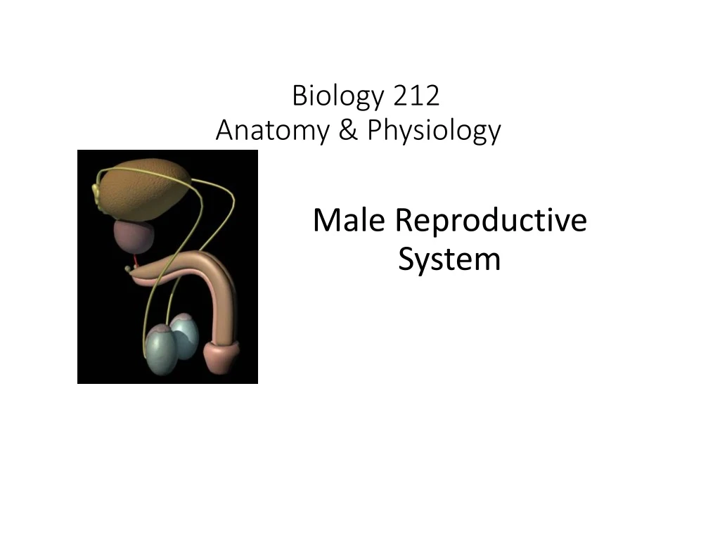
Biology 212Anatomy & Physiology I Male Reproductive System
Human Reproductive System: - Begins developing in 4th week of embryonic development. Remains “sexually indifferent” until 7th week in male 8th week in female
Both sexes: Gonads (ovaries & testes) remain inactive until puberty, when adenohypophysis (anterior pituitary) stimulates: production of gametes (sperm & oocytes) secretion of gonadal hormones
Cremaster Muscle (part of external oblique muscle) Scrotum
Spermatic Cord containing Vas Deferens Testicular Nerve, Artery & Vein Plexus Epididymis Testis
Testis or Testicle ~ 4 cm x 2 cm x 2 cm Surrounded by dense irregular connective tissue: Tunica Albuginea outside of which is pouch of peritoneum “dragged” into scrotum: Tunica Vaginalis
Testis or Testicle Each testis divided into approximately 250 Lobules by connective tissue Septa
Testis or Testicle Each testis divided into approximately 250 Lobules Each lobule contains one to four coiled Seminiferous Tubules where Sperm are produced
Testis or Testicle Each lobule contains one to four coiled Seminiferous Tubules where Sperm are produced
Sustentacular Cells Testis or Testicle Spermatogonia Primary Spermatocytes Secondary Spermatocytes Spermatids Sperm
Testis or Testicle Sperm production (spermatogenesis) by seminiferous tubules under control of follicle stimulating hormone produced by pituitary gland. Days for completion of Speciesspermatogenesis Human 64 Bull 54 Ram 49 Boar 34 Rat 48
Testis or Testicle Cellsbetween seminiferous = tubules Interstitial cells Secrete testosterone under stimulation by luteinizing hormone (interstitial cell stimulating hormone) produced by pituitary gland
Testis or Testicle Sperm pass from seminiferous tubule into tubulus rectus
Testis or Testicle Sperm pass from seminiferous tubule into tubulus rectus then into rete testis
Sperm pass from seminiferous tubule into tubulus rectus then into rete testis through efferent ductules
Sperm pass from seminiferous tubule into tubulus rectus then into rete testis through efferent ductules to epididymis
Epididymis Long, coiled tube In scrotum Posterior to testis Sperm take weeks to pass through epididymis where they mature: become fertile become motile and are stored until ejaculation Debris also removed in epididymis
Sperm from epididymis pass into vas deferens or ductus deferens which enters spermatic cord
Spermatic cord: Vas Deferens Testicular artery Testicular veins Autonomic nerves Cremaster muscle from external oblique Tunica vaginalis extension of peritoneum All wrapped in External Spermatic Fascia
Spermatic cord: Passes through inguinal canal into abdomen
Within abdomen, vas deferens passes to posterolateral side of bladder Lateral View
Within abdomen are two accessory glands: the seminal vesicles and the prostate Posterior View
Seminal Vesicles Paired, posterior to bladder
Seminal Vesicles Paired, posterior to bladder Join with ampulla of vas deferens to form ejaculatory duct
Seminal Vesicles Paired, posterior to bladder Join with ampulla of vas deferens to form ejaculatory duct Produces ~60% of fluid in semen Contains nutrients for sperm chemicals to decrease viscosity of cervical mucous & stimulate peristalsis of uterus
Prostate Inferior to bladder Surrounds ejaculatory ducts and proximal part of urethra
Prostate Inferior to bladder Surrounds ejaculatory ducts and proximal part of urethra Produces ~30% of fluid in semen Contains chemicals to enhance sperm motility & clot & then liquify semen & neutralize acidity of vagina
Semen = mixture of sperm from vas deferens seminal vesicle fluid prostate fluid enters urethra which passes through the penis
Penis Three erectile bodies Two corpora cavernosa One corpus spongiosum surrounding urethra
Penis: Shaft Bulb or Root Glans Prepuce (foreskin)
Erection: Parasympathetic stimuli cause arterioles in three erectile bodies (corpus spongiosum and corpora cavernosa) to dilate. The erectile bodies fill with blood and swell. This also presses on veins of the penis, restricting blood flow out of the penis.
Ejaculation: Physical stimulation of penis sends afferent nerve signals to lumbar spinal cord. Reflexive efferent nerve signals: 1) Sympathetic, causing: Constriction of bladder sphincters Peristaltic contraction of epididymis, vas deferens, & ejaculatory ducts move sperm into urethra Contraction of seminal vesicles and prostate move their secretions into urethra
Ejaculation: Physical stimulation of penis sends afferent nerve signals to lumbar spinal cord. Reflexive efferent nerve signals: 2) Somatic, causing: Contraction of skeletal muscles along bulb of penis force semen along urethra and out external urethral orifice
External Urethral Orifice (through Corpus Spongiousum) Urethra (through Prostate) At ejaculation Ejaculatory Duct Seminal Vesicles Ductus Deferens Constant Epididymis Efferent Ductules Tubulus Rectus Rete Testis Seminiferous tubules
Semen: Mixture of spermatozoa seminal vesicle fluid prostatic fluid Normal ejaculate: 4 – 6 ml 50 – 100 million sperm per ml Greatest in adolescence & young adults Decrease with age
Your assignment for tonight: Identify scrotum & testicles Identify location of epididymis, vas deferens, inguinal canal, prostate, seminal vesicles, urethra Identify corpora cavernosum & corpus spongeosum in both bulb and shaft of erect penis Observe clotting & liquification of freshly ejaculated semen