Download
1 / 23
230 likes | 254 Views
Explore various necrotic lesions and infections found in fish spleens, such as haemorrhage and viral diseases like HVHN and Iridovirus, affecting their architecture. Learn about unique patterns and characteristics in fish spleens.

E N D
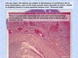
Like any organ, fish spleens are subject to disturbances of architecture due to local catastrophes, such as this large necrotic lesion (possibly an infarct, related to stripping trauma) in a post-spawned rainbow trout. (Scanned slide). There is considerable haemosiderosis and more recent haemorrhage surrounding the older lesion, but the remaining areas of spleen appear relatively normal. Liver Kidney Spleen Sp 40 x 4o Sp 40
This is a low power view of an old slide of a Greenback flounder spleen. Note the large irregular pale areas. .. Which at higher magnification are obviously necrotic foci with a little peripheral reaction. S12x 10o S12x 4o
Another area from this spleen shows focal fibrin deposits within some necrotic foci, and ….. We have seen this pattern before in the kidney. Can you recall the organism? Aeromonas salmonicida (in this case an atypical strain), the hall-marks being a sticky non-motile bacteria (resulting in clumps) and necrotizing toxins. … peripheral basophilic foci Fibrin Sp12 x 40o Sp12 x 20o S12x 10o
Focal spleen reactions & disturbances of architecture 2. Viral diseases
This Goldfish spleen also shows irregular mottled pale areas. These are more widespread than in the previous example. (Ignore the old granulomas: they are having no noticeable impact.) Higher magnification shows loss of architecture, with virtually no recognizable ellipsoid complexes and few recognizable cells, other than some remaining lymphoid tissue and blood cells. S13 x 10o S13 x 4o
With x40 objective, many cell fragments are visible, confirming the necrotic nature of the lesions, as well as many cells with swollen nuclei and generally swollen vacuolated cytoplasm. Note also the large lightly melanised cells of similar size – apparently the phagocytosed remnants of these enlarged abnormal cells. S13 x 40o
Another field (also x40o) shows occasional enlarged nuclei with a haloed eosinophilic inclusion (Cowdry Type A). S13 x 40o
Two fields under oil emersion (x100o) showing a) the appearance of the Cowdry Type inclusion b) the glassy fine contents of the other enlarged nuclei. Both show margination of host chromatin and both are distended with a herpes virus. which has already destroyed most of the spleen cells. Quiz: which cells do you think have been affected? a b S13 x 100o
Goldfish herpes virus The virus in these fish is Cyprinid herpesvirus 2 (CyHV-2), which is a pathogen of the goldfish Carassius auratus auratus L. This virus causes herpesviral hematopoietic necrosis (HVHN) disease, resulting in anemia with high mortality. Differs from related viruses: CyHV-1 (“carp pox” or fish papilloma virus) and the OIE listed CyHV-3 (koi herpes virus, also known as carp interstitial nephritis and gill necrosis virus) Although Cowdry type A inclusions [eosiniphilic inclusions surrounded by a halo] are typical of mammal herpes viruses, they are generally rare in fish virus disease, the less compact “glassy” nuclear distention being more common. Goldfish herpes virus was apparently absent, or at very low levels in Australian stocks, though recent incursions have been seen. (This case was a single fish from 1992 that had been isolated in current tank for some years - earlier history unknown.) Reference: Stephens FJ, Raidal SR, Jones B (2004) Haematopoietic necrosis in a goldfish (Carassius auratus) associated with an agent morphologically similar to herpesvirus. Aust Vet J 82:167–169
Recognize the organ? x 100o This is the head kidney of another Goldfish from a more recent outbreak of this virus disease (there was no spleen in section). Did you recognize the thyroid follicles? As the kidney is the other major haematopoietic organ, it is not surprising that this too contains large pale foci of necrosis. Note similar inclusions to those in the spleen x 40o x 20o x 10o x 4o
Spleen of a Dwarf Gourami. The regular architecture is also disrupted, though with less overt necrosis. The architecture is generally still present, but disrupted by unusual large cells. These cells are much larger than normal host cells (but they are host cells). As both nucleus and cytoplasm are very large, they are not infrequently mistaken for parasites, but as with the herpes virus above the enlargement is due to expansion by virus particles. S14 x 10o. S14 x 4o. S14 x 40o
Iridovirus of Gourami - & other fish • We have seen iridovirus infection previously in the kidney – (e g EHN – epizootic haematopoietic necrosis virus) but that involved overt necrosis, which is not an obvious feature of this case. • This highlights the 2 distinct patterns of fish iridovirus infection: • A) as in EHN: the cell becomes filled with viral particles, apparently very quickly, based on the outbreak pattern, resulting in cell rupture (ranaviruses). (Pathogenesis of the herpes virus above is similar.) • Or • B) as in Gourami iridovirus: virus continues to multiply, expanding but not initially rupturing the cells. (megalocytiviruses ) [Note spelling – megalocytovirus is the mammalian herpes virus version.] • Both groups of iridovirus contain internationally important (and reportable) pathogens - eg the megalocytivirus Red Sea Bream Iridovirus (which is known to affect over 30 species of marine fish): this and related strains cause significant diseases in farmed marine finfish in Asia. Megalocytiviruses also affect a range of ornamental fish species including gouramis (Colisa spp. and Trichogaster spp.) swordtails (Xiphophorus helleri), platies (Xiphophorus maculatus), mollies (Poecilia latipinna), angelfish (Pterophyllumscalare), orange chromides (Etroplus maculatus) and others. • There appears to be limited host specificity, but the number of related viruses or strains is not known. [We have seen concurrent infection of post quarantines stocks of dwarf gourami and angel fish.] • Megalocytiviruses from imported Gouramis has been implicated as a cause of severe mortality in cultured Murray Cod (>90% mortality of fingerlings), and experimentally transmitted to this species. It is closely related to Infectious Spleen and Kidney Necrosis Virus (ISKNV) which causes mass mortality in mandarin fish, a freshwater food fish cultured in China. (J. Go, R. Whittington / Aquaculture 258 (2006) 140–149) • It is important to establish the relationship of viruses found in aquarium fish to internationally recognised pathogens, and to consider cross-species infections. Investigation of the relationships of this virus (which enters Australia frequently with infected aquarium fish) is continuing (FRDC 2007/007, Whittington)
Some more examples from this fish: note that the endothelial cells also appear to be affected. S14 x 20o S14 x 40o
Note the variation in color, presumably reflecting the amount of virus. (Virus particles contain a high proportion of nucleic acid, and therefore stain a slightly basophilic purple, as for host nucleic acid.) S14 x 40o
.. And also, in this case, within the glomerular capillaries. Megalicytiviruses affect many species and their relationships are still unfolding. As both haematopoitic tissue and endothelial cells can be affected, lesions may be seen in a wide range of organs, but are most abundant in the haematopoietic tissue of kidney and spleen. This slide shows similar iridovirus induced hyperplastic cells in the highly vascular choroid complex of the eye of a Swordtail (another affected aquarium fish species). S14 x 40o
Focal spleen reactions & disturbances of architecture 3. Contained or partially contained bacterial diseases (post septicaemia) Granulomas (contained) bacterial and parasitic disease
This spleen of an Atlantic salmon infected with the Tasmanian rickiettsia-like organism (RLO) shows a widespread and irregular pattern of pallor, as well as the single large multicentric “bulls-eye” necrotic lesion (which, while indicative of rickettsia infections, is relatively rare).
This is a similar spleen (Atlantic salmon with RLO) showing the widespread scattered intracellular organisms, with little regularity with regard to location. This will include those multiplying within the host cells, as well as some being killed following phagocytosis (depending on the stage of infection). Note the early pigment (pale orange-red), as well as apparently live rickettsia organisms within this cell (arrow). And the more developed pigment, plus apparently live organisms, in this cell, suggesting some but not all the bacteria have been killed. S31 x 100o S31 x 40o
This spleen is also of Atlantic salmon with RLO, but apparently of longer duration, with a very marked spleen reaction. .. which in this case includes a Splendore-Hoeppli reaction (which we saw in the kidney as a response to Nocardia). Sx x 40o Sx x 4o
As one would expect of a filtration organ with major reticular-macrophage functional units, granulomas (formed against organisms that are phagocytosed or surrounded but not destroyed) and a common finding in the spleen of fish. Typical gross appearance of florid granulomatous lesions in the spleen, in this case a Chilean Atlantic salmon with Bacterial Kidney Disease (Renibacterium salmoninarum). [We saw similar granulomas in the kidney presentations.]
Rainbow trout with several large reactions within a generally reactive spleen. Which at higher power are also seen to be granulomas. x 10o Sx061573-7x 4o Sx x 20o
Another field from this Rainbow trout, showing giant cells and the nature of inciting cause for the granulomatous reaction. Can you recall the name of the organis? (Ichthyophonus hoferi) Although this course is using a systematic approach to the pathology of fish, organisms affecting one organ are likely to affect other organs with the same type of tissue. Sx x 20o Sx x 10o
fin These are explanatory presentations, to understand the nature of fish reactions and tissue appearance. General Reference to further consider the range of pathogens that may affect the spleen (or other organs) Ferguson, Hugh W. (Ed) 2006. Systemic Pathology of Fish (Second Edition). Scotian Press, London. (367 pp)