Download
1 / 33
330 likes | 365 Views
Explore the chambers of the heart and external features of the circulatory system. Learn about heart structure, pulmonary and systemic circulation, and vital facts about this essential system.

E N D
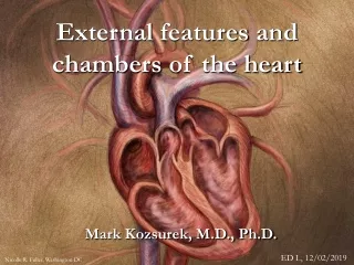
External features and chambersof the heart Mark Kozsurek, M.D., Ph.D. ED I., 12/02/2019 Nicolle R. Fuller, Washington DC
Unicellular organisms (e.g. Paramecium on the photo above) All the physiological functions (food-intake, excretion, movement, reproduction, etc.) are performed by one single cell. And all the cells belonging to the same species have the same morphological and physiological properties.
Locomotor system Digestive system Respiratory system Urogenital system Nervous system Circulatory system Multicellular organisms: groups of cells are getting segregated and specialized for a function, thus, organs/organ systems are formed (differentiation). (and several more systems…) The circulatory system is the transport pathway connecting all the organ systems.
The circulatory system LUNG Pulmonary/respiratory circulation HEART Systemic/general circulation BODY
Facts about the circulatory system • The circulatory system is the first system that appears in the embryo. Heart primordium starts beating by the end of the third week! • Deseases related to the heart or involving the circulatory system are the most frequent causes of death all over the world!
The center of the circulatory system is the heart weighting approx. 4 g/body mass kg, which means roughly 280-320g in an adult. • As the systemic and respiratory circulations are connected in series, the two circuits carry the same volume of blood. Calculating with a resting heart rate of 72 beat/min and 70-80 ml of blood forwarded by each contraction, 5.0-5.5 litres of blood are kept in motion every minute both in the systemic and respiratory circulation. • The resistance and the blood pressure is much higher in the systemic circulation (120/80 Hgmm vs. 24/9 Hgmm). As the left half of the heart has to work against a higher resistance, its wall is thicker, and the increased mass of cardiac muscle requires more nutrients and oxygen. Due to this the left half of the heart is more vulnerable and is more frequently involved in diseases.
A) Where is the heart? • Heart rests on the diaphragm in the anterior-inferior mediastinum.
Mediastinum posterior mediastinum anterior mediastinum supracardiac mediastinum thymus, great vessels cardiac mediastinum heart Mediastinum is an area within the thoracic cage not occupied by the lungs.
= sternocostal surface = posterior surface = inferior surface
1. Sternocostal surface 2/3 right ventricle 1/3 left ventricle auricles
2/3 right ventricle 1/3 left ventricle auricles
1. Sternocostal surface • The anterior interventricular sulcus with the anterior interventricular branch of the left coronary artery and the accompanying great cardiac vein represents the border between the two ventricles. • Coronary sulcus separating the atria from the ventricles is almost perpendicular to the anterior interventricular sulcus. It is not a complet ring as it is interrupted anteriorly by the root of the pulmonary trunk. In its right half the coronary sulcus contains the right coronary artery and the small cardiac vein.
2. Diaphragmatic surface 2/5 right ventricle 3/5 left ventricle right atrium at the IVC
2/5 right ventricle 3/5 left ventricle right atrium at the IVC
2. Diaphragmatic surface • The posterior interventricular sulcus containing the posterior interventricular branch of the right coronary artery and the accompanying middle cardiac vein is the landmark between the left and right ventricles. • Coronary sulcus is filled almost along its whole length with the coronary sinus collecting the majority of the veinous blood of the heart. Posteriorly, in the left-superior portion of the coronary sulcus the circumflex branch of the left coronary can be found, while in the right-inferior part of the groove the right coronary artery with the small cardiac vein is visible.
3. Posteriorsurface (base) left atrium
3. Posteriorsurface (base) • Almost exclusively formed by the left atrium. • Tight relation between the posterior wall of the left atrium and the esophagus (splinter, endoscopy, esophagus cancer) Sobotta
4. Left and right pulmonary surfaces Areas directed toward the lungs, some terminologies do not even mention them as they might be considered as the lateral transitional zones among the previously discussed three big surfaces!
Topography of the great vessels Anterior (A) and posterior (B) aspect of the heart and the great vessels.
Right atrium Eustachian-valve Thebesian-valve
The right atrium develops from two different sources and due to this consists of two parts. The posterior region termed sinus venarum has smooth wall and receives the openings of the superior and inferior vena cava as well as the opening of the coronary sinus. The wall of the anterior portion, mainly represented by the right auricle has rough wall due to the presence of the comb-like pectinate muscles. The two parts merge along the crista terminalis inside, while on the external surface the sulcus terminalis can be found in the same position. Note that the pectinate muscles and the crista terminalis are perpendicular to each other! • Find the valve of the inferior vena cava (Eustachian valve) and the valve of the coronary sinus (Thebesian valve) at the orifices of the corresponding veins! • Identify the fossa ovalis and its limbus on the interatrial septum!
Right ventricle Supraventricular crest „moderator band”
The right ventricle is a V-shaped chamber in which the inflow and the outflow parts (the latter is also termed conus arteriosus) are separated by the supraventricular crest. • The interior of the right ventricle is rough due to the irregular trabeculae carneae. Constant structures that can be found in the right ventricle are the anterior papillary muscle and the septomarginal trabecula (or moderator band). Posterior papillary muscle is negligible or is completly absent. The tree leaflets of the tricuspid valve are anchored through the chordae tendineae to the papillary muscles (anterior and posterior cusps) or directly to the wall (septal cusp).
A cuboidal chamber of the heart where the four corners are determined by the entrance ot the four pulmonary veins. Its interior is smooth walled (as it develops by the incorporation of the above mentioned pulmonary veins), pectinate muscles making the wall irregularly rough are restricted into the left auricle. • The valve of foramen ovale is sometimes visible, but is usually completly fused with the interatrial septum.
The internal surface is trabeculated (trabeculae carneae) just like in the right ventricle. There are two prominent structures: the anterior and the posterior papillary muscle. Note that both the leaflets of the mitral valve are attached onto both of the papillary muscles! • Inflow and outflow paths in the left ventricle are only separated by a fibrous structure: the anterior cusp of the mitral valve.
ant. cusp of mitral valve supraventricular crest
Sobotta left ventricle right ventricle Sobotta
http://punkagothic1.deviantart.com/art/my-Sullen-heart-273623478http://punkagothic1.deviantart.com/art/my-Sullen-heart-273623478 Oh, Heart Oh heart, oh heart, why must you beat? Oh heart, oh heart, why must you weep? Oh heart, oh heart, why must you hide? Oh heart, oh heart, what lies inside? Oh heart, oh heart, why must you cry? Oh heart, oh heart, why do you despise? Oh heart, oh heart, why are you weak? Oh heart, oh heart, why can't you speak? Oh heart, oh heart, why must you burn? Oh heart, oh heart, why can't you learn? Oh heart, oh heart, what went wrong? Oh, heart, oh heart, can't you be strong? Oh heart, oh heart, why are you blue? Oh heart, oh heart, where are you? Whitney Albright Thank you for your attention!