Download
1 / 13
130 likes | 152 Views
This clinical case study focuses on a male patient who presented with a swollen and painful right lower limb, along with skin damage. The case highlights the importance of accurate diagnosis in identifying the underlying cause of the swelling and provides insights into venous obstruction and the rare occurrence of a lipoma causing venous compression. The patient's morbid obesity and previous history of deep venous thrombosis are also discussed.

E N D
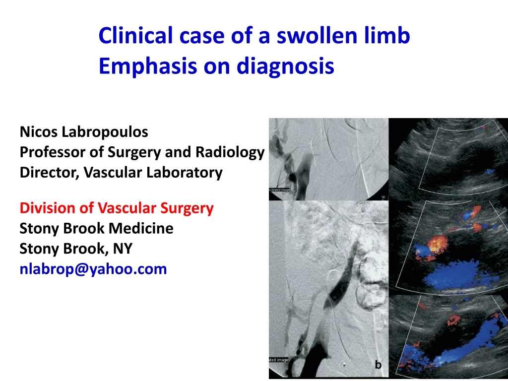
Clinical case of a swollen limb Emphasis on diagnosis Nicos LabropoulosProfessor of Surgery and RadiologyDirector, Vascular LaboratoryDivision of Vascular SurgeryStony Brook MedicineStony Brook, NYnlabrop@yahoo.com
Male 49y morbidly obese presented with RT LE swelling, pain and skin damage Right thigh circumference 70cm and the left 54cm. Diabetes, hypertension and previous RT LE DVT No history of surgery, trauma, or long distance travel
70cm 54cm Bilateral swelling RT>LT RT Extending to foot and toes Circumferential skin changes Healed ulcer
2001Initial duplex ultrasound showed isolated CFV thrombosis measuring 9.6mm. The patient was treated with anticoagulation. 2003 Pain and worsening of the swelling DU showed CFV thrombosis measuring 22mm
Right LE ultrasound CFV Duplex ultrasound in 2003 showed persistent CFV occlusion measuring 22mm, which progressed from 9.6mmin 2001.
Later, he developed a venous ulcer that was managed with inelastic compression bandages. DU showed no deep or superficial reflux, with isolated CFV occlusion (diameter of 30 mm). Suspicion for underlying iliac venous outflow obstruction prompted a CTV showing a mass with cystic characteristics adjacent to CFV measuring 30mm.
Right LE ultrasound and CTV 2008persistent CFV occlusion of the common femoral vein and increase in size to 30mm. Cystic appearing mass compressing the CFV and displacing the femoral vessels
The mass, which was previously thought to be a thrombosed CFV, was compressing the vein and causing venous outflow obstruction. The patient refused any surgical intervention and was lost to follow-up. He finally returned with worsening symptoms, and repeat venous DU and a venogram were performed.
Right LE ultrasound and venogram Duplex ultrasound and venography with minimal flow in supine position. Distal augmentation demonstrates patency of the CFV. Pressure gradient 15mmHg across the occlusion. Differential diagnosis -Vein wall tumor -Adventitial cyst
An encapsulated mass was identified, which was compressing the underlying common femoral vein. An extravascular lipoma was released once the capsule was opened.
Underlying common femoral vein following removal of lipoma. Low power view of lipoma, x40 magnificationdemonstrated varying sizes of normal adipocytes.
Postoperatively, the patient developed superficial fat necrosis, which required wound debridement and VAC therapy. At 3-month follow-up, the thigh circumference in the RT thigh was 58cm, his edema improved, the leg ulcer healed, and DU showed a patent CFV. Lipoma is a rare cause of venous obstruction with <10 cases reported and at least half are intravenous.
Morbid obesity can explain the bilateral lower limb swelling. RT thigh and leg were still bigger than the left after the treatment. The right limb due to chronic venous obstruction may have developed lymphedema.