Embryology – 2 nd
Embryology – 2 nd. Dr. Khaldoun Darwich. Blastocyst stage. The trophoblast layer gives rise to important prenatal support tissues The embryoblast layer gives rise to the embryo during the next prenatal period. Developmental Disturbances during Meiosis.


Embryology – 2 nd
E N D
Presentation Transcript
Embryology – 2nd Dr. KhaldounDarwich
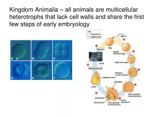
Blastocyst stage The trophoblast layer gives rise to important prenatal support tissues The embryoblast layer gives rise to the embryo during the next prenatal period
Developmental Disturbances during Meiosis If any disturbances occur in meiosis during fertilization major congenital malformations result from the chromosomal abnormality
Developmental Disturbances during Meiosis In Down syndrome or Trisomy 21, an extra chromosome number 21 is present after meiotic division
Down syndrome A child with this syndrome has a: flat broad face with wide set eyes , a flat bridged nose , epicanthic folds , oblique eyelid fissures , a furrowed lower lip , fissures of the tongue , hypertrophy of the lingual papillae , and other defects .
Down syndrome An affected child can have various levels of mental retardation
Down syndrome Children with Down syndrome may have increased levels of : periodontal disease and fewer and abnormally shaped teeth , presenting challenges to oral hygiene care. The arched palate and poor use of tongue muscles lead to an open mouth position and protrusion of the normal-sized tongue.Therefore articulate speech is often difficult.
Ectopic pregnancy Implantation may occur outside the uterus, a condition called ectopic pregnancy. Most of these occur in the fallopian tube. This disturbance has, several causes but is usually related to factors that delay or prevent transport of the dividing zygote to the uterus, such as scarred uterine tubes due to pelvic inflammatory disease. Tubal pregnancies usually Rupture, causing loss of the embryo and threatening the life of the pregnant woman.
EMBRYONIC PERIOD OF PRENATAL DEVELOPMENT The second period, the embryonic period of prenatal development extends from the beginning of the second week to the end of the eighth week.
EMBRYONIC PERIOD OF PRENATAL DEVELOPMENT the physiological processesoccuring during this period include : Induction : Interaction between embryological cells Proliferation : Controlled cellular growth and accumulation of byproducts Differentiation : Change in identical embryonic cells to become distinct structurally and functionally Morphogenesis : Development of specific tissue morphology or differing form due to embryonic cell migration and inductive interactions Maturation : Attainment of adult function and size due to proliferation, differentiation, and morphogenesis
EMBRYONIC PERIOD OF PRENATAL DEVELOPMENT These processes cause the structure of the implanted blastocyst to become an embryo These physiological processes also allow the teeth and associated structures, as well as other organ structures, to develop in the embryo
Induction The first physiological process involved in the beginning of most embryological development It is the interaction between embryological cells. Just what triggers cells to develop into structures from cellular interactions is poorly understood, but many problems can result from a failure of induction, leading to failure of initiation of certain embryological structures. Induction can also occur in the later stages of development.
Proliferation It means controlled levels of cellular growth that is present during most of embryological development. Finally, growth also occurs because of an accumulation of cellular byproducts
Proliferation Growth may be interstitial growthwhich occurs from deep within a tissue or organ (soft tissue growth). or appositional growth, in which a tissue enlarges its size by the addition of layers on the outside of a structure (Hard tissues such as mature bone or dental tissues) . Some tissues such as cartilage and growing bone tissue use both types of growth to attain their natural size .
Differentiation In the process of differentiation, a change occurs in the embryonic cells, which are identical genetically but become quite distinct structurally and functionally. Thus, cells that perform specialized functions are formed by differentiation during the embryonic period. Although these functions are minimal at this time, the beginnings of all major tissues, organs, and organ systems are formed during this period from these specialized cells.
Differentiation Differentiation occurs at various rates in the embryo. Cytodifferentiation: is the development of different cell types. Histodifferentiation: is the development of different tissues. Morphodifferentiation: is the development of the differing form, or morphology, for an organ or system.
Morphogenesis morphogenesis: process of development of specific tissue morphology. Morphogenesis is due to the migration of embryonic cells and inductive interactions of those cells. induction continues to occur during the embryonic period because the new varieties of cells interact with each other, producing an increasingly complex organism.
Maturation Finally, the physiological process of maturation of the tissues and organs begins during the embryological period and continues during the later fetal period. It is important to note that the physiological process of maturation of the individual tissues and organs also involves proliferation, differentiation, and morphogenesis. Thus, maturation is not the attainment of just the correct adult size but also the correct adult form and function of tissues and organs.
An embryo is recognizably human at the end of the embryonic period, or the end of the eighth week of prenatal development. • the major events of the second, third, and fourth weeks of the embryonic period will be next discussed.
Second WeekOf Prenatal Development During the second week of prenatal development, within the embryonic period, the implanted blastocyst grows by increased proliferation of the embryonic cells, as well as cellular morphogenesis and differentiation. The increased number of embryonic cells creates the embryonic cell layers, or germ layers within the blastocyst. A bilaminar embryonic discis eventually developed from the blastocyst. The bilaminar embryonic disc appears as a flattened, essentially circular plate of bilayered cells.
Week 2 • Bilaminar embryonic disc
Week 2 • Bilaminar embryonic disc
2nd Week :the Bilaminarembryonic disc This bilaminar disc has a superior epiblast layer and an inferior hypoblast layer Theepiblast layer is composed of high columnar cells, and the hypoblast layer is composed of small cuboidal cells. The bilaminar disc develops into the embryo as prenatal development continues.
2nd Week :the Bilaminar embryonic disc After its creation, the bilaminar disc is suspended in the uterus's endometrium between two fluid-filled cavities. the amniotic cavity, which faces the epiblast layer, and the yolk sac, which faces the hypoblast layer. The yolk sac serves as initial nourishment for the embryonic disc.
Week 2 • Bilaminar embryonic disc
2nd Week :the Bilaminar embryonic disc Later, the placenta, a prenatal organ that joins the pregnant woman and developing embryo, develops from the interactions of the trophoblast layer and endometrial tissues. The formation of the placenta and the developing umbilical circulation permit selective exchange of soluble blood-borne substances between the woman and the embryo.
Week 2 • Bilaminar embryonic disc
Third Week Of Prenatal Development During the beginning of the third week of prenatal development, the Primitive Streak forms within the bilaminar disc This furrowed, rod-shaped thickening in the middle of the disc occurs because of increased proliferation of cells in the midline area. The primitive streak causes the disc to have bilateral symmetry, with a right half and left half. Most of the further development of each half mirrors the other half of the embryo.
3rd Week the Primitive Streak • Embryonic bilaminar disc • Epiblast layer — • Hypoblast layer- • Yolk sac/ • Figure 3-6 • The bilaminar embryonic disc and formation of the primitive streak, with the resulting bilateral symmetry. • Embryonic bilaminar disc • Epiblast layer — • Hypoblast layer- • Yolk sac/
Third Week Of Prenatal Development During the start of this third week, some cells from the epiblast layer move or migrate toward the hypoblast layer in the area of the primitive streak. These migratory cells locate in the middle between the epiblast and hypoblast layers, becoming mesenchyme, an embryonic connective tissue. Mesenchymal cells have the potential to proliferate and differentiate into diverse types of connective tissue forming cells (e.g., fibroblasts, chondroblasts, and osteoblasts). Some of this tissue creates a new embryonic layer, called the mesoderm
Week 3 Bilaminar to trilaminar disc
Week 3 Bilaminar to trilaminar disc
Third Week Of Prenatal Development With three layers present, the bilaminar disc has become thickened into a Trilaminar embryonic disc. Thus, the Trilaminar disc has three embryonic layers, or germ layers. With the creation of this new embryonic layer (Mesoderm), the epiblast layer is now considered ectoderm and the hypoblast layer is endoderm.
Third Week Of Prenatal Development Within the trilaminar disc, each embryonic layer is distinct from the others and thus gives rise to specific tissues. The ectoderm gives rise to the epidermis of the skin, the nervous system, and other structures. The mesoderm gives rise to muscle coats, connective tissues, vessels supplying tissues and organs, and other tissues. The endoderm gives rise to the epithelial linings of the respiratory passages and digestive tract, including some glandular organ cells.
Third Week Of Prenatal Development The trilaminar disc now has a cephalic end, or head end. At the cephalic end, the oropharyngeal membrane, forms. This membrane consists of only ectoderm externally and endoderm internally. This membrane is the location of the future primitive mouth of the embryo and thus the beginning of the digestive tract.
Third Week Of Prenatal Development The disc also has a caudal end , or tail end . At the caudal end, the cloacal membrane forms. This is the location of the future anus, the terminal end of the digestive tract. Similar to the oropharyngeal membrane, the cloacal membrane consists of only two embryonic layers, without any mesoderm.
Third Week Of Prenatal Development During the later part of the third week of prenatal development, the central nervous system (CNS) begins to develop in the embryo Many steps occur during this week to form the beginnings of the spinal cord and brain
Third Week Of Prenatal Development (CNS) • First, a specialized group of cells differentiates from the ectoderm. • These cells are the neuroectoderm, and they are localized to the neural plate of the embryo. • The neural plate is a band of cells that extends the length of the embryo, from the cephalic end to the caudal end. • This plate undergoes further growth and thickening, which cause it to deepen and invaginate centrally, forming the neural groove.
Third Week Of Prenatal Development Near the end of the third week, the neural groove deepens further and is surrounded by the neural folds As further growth of the neuroectodermoccurs, the neural folds meet superior to the neural groove and a neural tube is formed during the fourth week. The neural tube undergoes fusion at its most superior portion and forms the future spinal cord, as well as other neural tissues
Third Week Of Prenatal Development In addition, during the third week, another specialized group of cells, the neural crest cells, develop from neuroectoderm . These cells migrate from the crests of the neural folds and disperse within mesenchyme. These migrated cells are involved in development of many face and neck structures, such as the branchial arches.
Third Week Of Prenatal Development Many embryologists consider the neural crest cells to be a fourth embryonic layer In the future, these cells are involved in the formation of cranial supporting connective tissues, cartilage, bone, and certain dental tissues such as the pulp, the cementum, and periodontal ligament. Thus, neural crest cells are essential in the development of the face, neck and oral tissues
Third Week Of Prenatal Development By the end of the third week, the mesoderm additionally differentiates and begins to divide into paired cuboidal aggregates of cells called somites . These 38 paired blocks of mesoderm are located on each side of the developing midline portion of the central nervous system in the embryonic disc