Download
1 / 72
730 likes | 865 Views
Musculoskeletal Disorders. Megan McClintock, MS, RN Fall 2011. Skeletal Functions. Support and framework for body Protection of vital organs Assist with movement Blood cell production Mineral and salt storage. Structure. Bone Joints Cartilage Muscle Ligaments/Tendons Fascia Bursae.

E N D
Musculoskeletal Disorders Megan McClintock, MS, RN Fall 2011
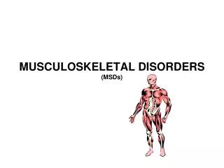
Skeletal Functions • Support and framework for body • Protection of vital organs • Assist with movement • Blood cell production • Mineral and salt storage
Structure Bone Joints Cartilage Muscle Ligaments/Tendons Fascia Bursae
Assessment - Subjective Gerontologic differences Past health history Medications Nutrition Occupation
Assessment - Objective Inspection Palpation Motion Muscle-Strength Testing Measurement Scoliosis Straight-leg raising test
Common Abnormalities • Table 62-6 • (pg 1577)
Diagnostic Studies Diskogram Myelogram DEXA Bone scan Arthroscopy Arthrocentesis EMG Duplex venous doppler SSEP
Labs Alkaline phosphatase Calcium Phosphorus RF ESR ANA Complement Uric acid CRP CK
Contusions • Soft tissue injury from blunt force • Overlying skin intact, but area becomes black and blue from localized hemorrhage • Usually only painful if palpated
Hematoma • Blood collection that occurs from torn blood vessel • Pain occurs as blood accumulates and places pressure on nerves • Pain occurs without palpation • Hematomas may burst or become infected
Strains • Overstretched tendons or overused muscles • Usually arise from twisting or wrenching movements • Acute – sudden, severe incapacitating pain with swelling • Chronic – repetitive movements; pain less severe but longer term (tennis elbow, runner’s knee)
Sprains • Ligament injuries • Grade 1 (mild) – small longitudinal ligament fiber separation • Grade 2 (moderate) - <100% of ligament is torn in cross-sectional direction. Function impaired • Grade 3 (severe) – ligament completely torn. Surgery required • Grade 4 (sprain fracture) – avulsion of bone fragment at site of ligament attachment
Interventions Prevent R – est I – ce C – ompress E – levate Analgesia as necessary After 24-48 hrs, warm moist heat
Subluxation/Dislocation • Bones are dislodged from normal positions within joints • Subluxation = partial dislocation • Joint capsule and ligaments damaged • Usually deformity at site • S/S: altered length of extremity, loss of function
Interventions Orthopedic emergency Assist with realignment Pain relief Restriction of movement Future activity restrictions
Fractures • Disruption in continuity of bone • Usually involves damage to surrounding soft tissue • S/S - pain, swelling, loss of function, deformity, abnormal mobility, bruising (also see pg 1591) • May be classified by severity and direction of fracture
Type of Fracture • Open (compound) • Closed (simple) • Incomplete • Complete • Displaced • Comminuted
Direction of Fracture • Transverse • Oblique • Spiral • Greenstick
Fracture Reduction Closed reduction ORIF (open reduction with internal fixation)
Fracture Repair Casting
Fracture Repair External fixation
Fracture Repair Internal fixation
Drugs Muscle relaxants Pain medications Tetanus prevention Antibiotics
Nutrition Ample protein Vitamins B, C, D Calcium Phosphorus Magnesium 2000-3000 mL/day of fluids High-fiber diet
Interventions • Assessment • Distal to the extremity • Neurovascular • Peripheral vascular • Peripheral neurologic • Prevention • Safety equipment • Elderly (also see pg 1584)
Interventions Pre-op skin prep Post-op neurovascular assessment Proper alignment & positioning Observe for bleeding, drainage Prevention of constipation Prevention of kidney stones Maintenance of cardiopulmonary system
Traction Interventions Inspect skin and pin sites carefully Pin site care Correct positioning ROM of unaffected joints Maintain traction at all times
Cast Care Interventions Handle a wet cast with palms only Support cast with pillows when wet Elevate at or above heart level Do not scratch skin with any objects Pad rough cast edges Can use cool air from hair dryer to help with itching Apply ice for first 24-36 hours Do not get cast wet
Fracture Complications • Direct • Infection • Inadequate bone union • Avascular necrosis • Indirect • Compartment syndrome • Venous thromboembolism (VTE) • Rhabdomyolisis • Fat embolism • Shock
Infection Venous Thromboembolism (VTE) • Esp. after hip fx, THA, total knee • Prevent – anticoagulants, SCDs, ROM to unaffected joints High incidence with open fx or soft tissue injury Need aggressive debridement
Compartment Syndrome Pressure that compromises neurovascular function Causes – restrictive dressings, edema S/S – Pain unrelieved by drugs and out of proportion – 1st, late is no pulses, paralysis, dark brown urine Tx – quick recognition, do NOT elevate, NO cold, fasciotomy
Fat Embolism Syndrome Systemic fat globules lodge in organs and tissues Risk with long bone, ribs, tibia, pelvis fx S/S – chest pain, tachypnea, dyspnea, change in mental status, hypoxia, petechiae on neck, chest, axilla, eyes, sense of impending doom Tx – early recognition!, reposition as little as possible, oxygen
Types of Fractures • Colles’ – wrist fx • Silver-fork deformity • Move thumb, fingers, shoulder • Humerus • Cx – radial nerve or brachial artery injury, frozen shoulder
Pelvic Fracture Can be life-threatening S/S – bruising on the abdomen, pelvis instability, swelling, tenderness Tx – Bed rest (few days to 6 weeks), may need traction, hip spica cast, ORIF, only turn when ordered by HCP
Hip Fracture 30% die within 1 year of injury S/S – external rotation, mm spasm, shortening of affected leg, severe pain Cx – nonunion, avascular necrosis, dislocation, arthritis Tx – surgery, may temp. use Buck’s traction
Hip Fracture Post-Op Care • Pillows/abductor splint between knees esp. when turning, avoid extreme hip flexion, don’t turn on affected side, OOB on first post-op day, in hospital for 3-4 days • Posterior approach • Table 63-11 (pg 1607) • No extremes in flexion • No putting on shoes, socks • No crossing the legs or feet • No low toilet seats • Precautions for 6 weeks • Anterior approach • Limited restrictions
Types of Fractures • Femoral Shaft • Can have lots of blood loss, risk of fat embolism • Tx – ORIF with traction after, hip spica cast • Tibia • Neurovascular assessment q 2 hrs x 48 hrs • Stable Vertebral • Logroll, orthotic devices, hard cervical collar • Vertebroplasty • Kyphoplasty
Facial Fractures • Impt to maintain patent airway, provide adequate ventilation • Assume that they have a cervical injury • Always have suction available • For jaw fractures: • Position pt on the side with head slightly elevated • Wire cutter/scissors at the bedside • Trach tray always available • NG tube decompression • Oral hygiene is impt • Protein supplements
Amputation • Pain is not a primary reason • Pre-op preparation • Post-op • Sterile technique for dressing changes • Immediate prosthesis vs delayed • Don’t sit in chair > 1 hr • Lie on abdomen 3-4 times/day • Residual limb bandaging • Table 63-14 (pg 1613)
Joint Procedures • Synovectomy • Removal of synovial membrane • Osteotomy • Remove a wedge of bone • Debridement • Removal of degenerative debris • Arthroplasty • Reconstruction or replacement of a joint
Total Hip Arthroplasty (THA) See notes from hip fracture Can’t drive or take tub bath for 4-6 weeks Knees must be kept apart Don’t cross legs Don’t twist to reach behind Quadriceps and hip muscle exercises High risk for thromboembolism No high-impact exercises/sports Usually stay in the hospital 3-5 days
Carpal Tunnel Syndrome Compression of the median nerve Women more likely to get S/S – thumb weakness, burning pain, numbness, parasthesia Tinel’s and Phalen’s sign http://tinyurl.com/cre5lf2 Tx – splints, rest, surgery
Rotator Cuff Injury Muscles that stabilize the humeral head and give ROM Cause – fall onto outstretched arm, repetitive overhead arm motion, heavy lifting S/S – shoulder weakness, pain, decreased ROM Drop arm test http://tinyurl.com/d2jq5jc Tx – RICE, corticosteroid injection, surgery
Meniscus Injury Occur with ligament sprains in a rotational force injury S/S – no edema (unless other injury), tenderness, pain, effusion in the joint, felt a “pop”, knee locks or gives way, MRI McMurray’s test http://tinyurl.com/cev9lx9 Tx – RICE, knee brace, arthroscopy, rehab starts quick Prevention – warm-up exercises