

E N D
Presentation Transcript
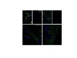
Figure 3. Immunofluorescence of in vitro infected myotubes using antibodies against the envelope and NS1 protein of DENV. Cultured human myotubes were infected with DENV (A,C,D,F) or exposed to mock infection (B,E) as described in methods, (A) the DENV structural protein E was visualized by indirect immunofluorescence (Green), the nuclei was stained with DAPI (Blue). (B) Mock infected myotubes are negative for the viral E protein. (C) A 100x image of the immunostaining against the E protein showing perinuclear localization. (D) Immunostaining against the non-structural viral protein NS1 (Green) and nuclei was stained with DAPI (Blue). (E) Mock infected myotubes are negative for the NS1 protein. (F) 100x image of the immunostaining against NS1 in infected myotubes. Scale bar 200 µm (A, B, D, E) and 20 µm (C and F).