Download
1 / 26
270 likes | 473 Views
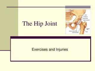
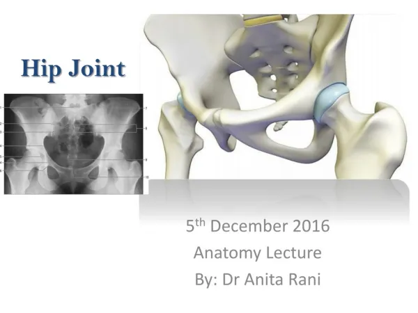
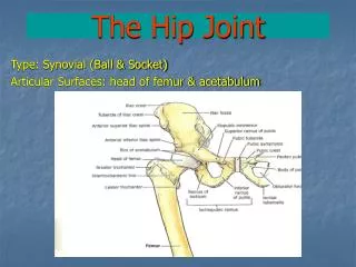
HIP JOINT. By: Dr. Mujahid Khan. Articulation. The hip joint is the articulation between the hemispherical head of femur and the cup shaped acetabulum of the hip bone The articular surface of the acetabulum is horseshoe shaped and is deficient inferiorly at the acetabular notch. Articulation.

E N D
HIP JOINT By: Dr. Mujahid Khan
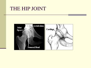
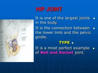
Articulation • The hip joint is the articulation between the hemispherical head of femur and the cup shaped acetabulum of the hip bone • The articular surface of the acetabulum is horseshoe shaped and is deficient inferiorly at the acetabular notch
Articulation • The cavity of acetabulum is deepened by the presence of a fibrocartilaginous rim called acetabular labrum • The labrum bridges across the acetabular notch and is here called the transverse acetabular ligament • The articular surfaces are covered with hyaline cartilage
Type & Capsule • It is a synovial ball and socket joint • The capsule encloses the joint and is attached to the acetabular labrum medially • Laterally it is attached to the intertrochanteric line of the femur in front and along the posterior aspect of the neck of the bone behind
Iliofemoral Ligaments • It is a strong, inverted Y-shaped ligament • Its base is attached to the anterior inferior iliac spine above • Below the two limbs of Y are attached to the upper and lower parts of the intertrochanteric line of the femur • The strong ligament prevents overextension during standing
Pubofemoral Ligament • It is a triangular ligament • The base of the ligament is attached to the superior ramus of the pubis • The apex is attached below to the lower part of the intertrochanteric line • This ligament limits extension and abduction
Ischiofemoral Ligament • It is a spiral shaped ligament • Attached to the body of the ischium near the acetabular margin • Fibers pass upward and laterally and attached to the greater trochanter • This ligament limits the extension
Transverse Acetabular Ligament • It is formed by the acetabular labrum as it bridges the acetabular notch • It converts the notch into a tunnel through which blood vessels and nerves enter the joint
Ligament of Head of Femur • It is flat and triangular ligament • It is attached by its apex to the pit on the head of the femur (fovea capitis) • Attached by its base to the transverse ligament and the margins of the acetabular notch • It lies within the joint and is ensheathed by synovial membrane
Synovial Membrane • The synovial membrane lines the capsule • It is attached to the margins of the articular surfaces • It covers the portion of the neck of the femur that lies within the joint capsule • It ensheathes the ligament of the head of the femur
Synovial Membrane • It covers the pad of fat contained in the acetabular fossa • A pouch of synovial membrane frequently protrudes through a gap in the anterior wall of the capsule • Forms the psoas bursa beneath the psoas tendon
Nerve Supply • Femoral nerve • Obturator nerve • Sciatic nerve • Nerve to the quadratus femoris
Movements • The hip joint has a wide range of movement but less so than the shoulder joint • Some of the movement has been sacrificed to provide strength and stability • The strength of the joint depends largely on the shape of the bones taking part in the articulation and on strong ligaments
Movements • When the knee is flexed, flexion is limited by the anterior surface of the thigh coming in contact with the anterior abdominal wall • When the knee is extended, flexion is limited by the tension of the hamstring muscles • Abduction is limited by the tension of the pubofemoral ligament
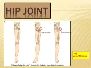
Movements • Adduction is limited by contact with the opposite limb and by the tension of the ligament of the head of the femur • Lateral rotation is limited by the tension in the iliofemoral and pubofemoral ligaments • Medial rotation is limited by the ischiofemoral ligament
Movements • Flexion: It is performed by the iliopsoas, rectus femoris, sartorius, also by adductor muscles • Extension: it is performed by the gluteus maximus and the hamstring muscles • Abduction: It is performed by the gluteus medius and minimus, assisted by sartorius, tensor fasciae latae, and piriformis
Movements • Adduction: It is performed by the adductor longus and brevis and the adductor fibers of the adductor magnus • Lateral rotation: It is performed by the piriformis, obturator internus and externus, superior and inferior gamelli • Medial rotation: It is performed by the anterior fibers of gluteus medius and gluteus minimus and the tensor fasciae latae • Circumduction: It is a combination of the previous movements
Movements • The extensor group of muscles is more powerful than the flexor group • The lateral rotators are more powerful than the medial rotators
Relations • Anteriorly: Iliopsoas, pectineus, and rectus femoris • Posteriorly: The obturator internus, the gamelli, and the quadratus femoris muscle separate the joint from sciatic nerve • Superiorly: Piriformis and gluteus minimus • Inferiorly: Obturator externus tendon