Download
1 / 1
10 likes | 155 Views
*. *. *. *. *. *. *. *. *. ABSTRACT. RESPONSE TO CHEMICALS

E N D
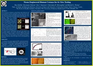
* * * * * * * * * ABSTRACT RESPONSE TO CHEMICALS Neuropeptides, such as substance P (SP) are released from nerve terminals in the epithelium. To elicit a functional response such as SP release, innervated corneas were treated with capsaicin or veratridine. Differential SP release was observed between test groups and controls (Fig. 7A), in a fashion similar to native nerve processes. We also showed that the presence of nerves in the TE cornea was able to protect the epithelium from chemical irritation. Innervated and non-innervated corneas were treated with a surfactant, and cell viability was examined with live/dead staining. There was significantly less cell death in innervated corneas (Fig. 7A, B). A We have previously tissue engineered a cell-based human corneal substitute that could serve as an alternative to animals in ocular irritancy testing. The reconstructed corneas resemble human eye bank corneas maintained in tissue culture. The constructs express early response genes, cytokines and extracellular matrix genes thought to play a role in ocular irritancy responses and wound healing. Results obtained using these models reflect in vivo irritancy potentials of test chemicals. The corneas also produced A B quantifiable changes in transparency corresponding to different degrees of cellular damage and irritation. However, while key structural and physiological features of natural corneas were reproduced, no indications of sensitivity and pain were obtained. We therefore added a nerve component to these tissue engineered corneas to allow for a more complete range of testing capabilities including neurotoxicity testing. Human corneal epithelial, stromal and endothelial cell lines with extended lifespans were used to construct a cornea around and within a stabilized collagen-GAG scaffold. With added nerve cells, the engineered corneas C no nerves B + nerves C D Green = live cells Red = dead cells Fig. 7 Fig. 3 CORNEAS FOR TRANSPLANTATION This corneal model was the starting point for the development of tough polymer composites for use in transplantation. The possibility for innervation was first tested in vitro. Confocal imaging confirmed the growth of nerves at different depths into the polymer (Fig. 8A). After implantation into corneas, the growth of nerves into the polymer was examined. Extensive innervation of the polymer was observed 3 months post-surgery (Fig. 8B) A ACTION POTENTIALS (APs) Electrophysiological recording from the corneal epithelium confirmed that the nerves were able to conduct lidocaine-sensitive action potentials that were evoked by stimulation of the ganglion. These APs (Fig. 4A) were similar to those recorded from native nerve endings both in configuration and amplitude8. show an innervation pattern resembling that of natural corneas. The in-growing nerves are excitable, propagate action potentials and show graded release of the neurotransmitter Substance P in response to neurotoxins. Differential wound healing rates in response to epithelial wounding are observed also. These innervated human corneas can therefore, in the future, be used as a more complete in vitro alternative to animals for ocular irritancy testing. Modifications to the basic technology developed to date allows for fabrication of other more complex tissues for testing applications and transplantation. similar to the natural cornea (inset). These penetrating nerve bundles branched to give finer nerve structures (Fig. 8C). This demonstrates the feasibility of developing transplantable artificial corneas that promote nerve regeneration following surgery. B A C B Fig. 4 Because of the close proximity of the electrodes, a large stimulus artifact was generated (Fig. 4B) that obscured the small AP (arrow). To isolate the AP, evoked responses were recorded before and after 50 mM lidocaine treatment. Subtracting the responses yielded isolated APs (Fig. 4A). Two controls were used: (a) innervated corneas with the recording electrode in an area without nerves and (b) corneas with no nerves. Responses of control corneas were measured with the same procedure and subtraction of pre- and post-lidocaine responses resulted in the loss of the response. P WOUND HEALING The loss of corneal innervation has been shown to slow wound healing2. To test if this was reproducible in our model, epithelial wounds (Fig. 5B) were made in constructs with and without nerves. Wound closure rates were then measured. During the first 18 hours post-wounding, innervated corneas showed faster wound closure (Fig. 5A). BrdU labelling, which measures proliferation, showed no increase in the mitotic index of epithelial cells over time, in either innervated or non-innervated corneas (Fig. 5C). This suggests that the faster initial healing of innervated corneas was likely due to faster migration of epithelial cells. These data are consistent with other studies that show increased proliferation beginning only 24 hours after injury9. A sclera C B cornea Fig. 1 Fig. 2 Fig. 5 EPITHELIAL CELL PROLIFERATION BrdU labelling was more intense in innervated constructs compared to non-innervated controls (Fig. 5C and 6A, B). This demonstrates that the presence of nerves in the cornea constructs promoted epithelial cell proliferation, and is consistent with historical in vivo rabbit studies3. + nerves no nerves • REFERENCES • C. R. Hicks et al., Prog. Retinal Eye Res. 19, 149 (2000). • R. W. Beuerman et al., Exp. Neurol. 69, 196 (1980). • K. Araki et al., Curr. Eye Res.13, 203 (1994). • T. Nishida et al., J. Cell Physiol.169, 159 (1996). • K. Araki-Sasaki et al., J. Cell Physiol.182, 189 (2000). • 6.M. Griffith et al., Science286, 2169 (1999). • L. J. Muller et al., Invest. Ophthalmol. Vis. Sci.37, 476 (1996). • J. A. Brock et al., J. Physiol.512, 211 (1998). • L. Gan et al., Acta. Ophthalmol. Scand. 79, 488 (2001). A B Fig. 6 Tissue Engineered Human Corneas for In Vitro Testing May Griffith1, Rosemarie Osborne2, Eric J. Suuronen1, Christopher R. McLaughlin1 and Mitchell A. Watsky3 1Dept. of Cellular and Molecular Medicine, University of Ottawa and University of Ottawa Eye Institute, Ottawa, Ontario, Canada; 2The Procter & Gamble Company, Cincinnati, OH; 3University of Tennessee Health Science Center, Memphis, TN. RESULTS and DISCUSSION • NERVE MORPHOLOGY • All corneal nerve fiber types were observed in our model. Using immunofluorescence with a nerve specific marker (anti-NF200), we were able to identify: • stromal nerves (fig. 3A) • a nerve plexus below the epithelium (Fig. 3B) • beaded (arrow) and smooth (arrowhead) fibers in • the epithelium (Fig. 3C). • These nerves were similar to their human counterparts (insets). • TEM showed nerve fibers invaginated epithelial cells (Fig. 3D; arrow). This suggests that cells receive direct innervation, as previously reported for human corneas7. Red = surface, green = -5 μm, blue = -15 μm OBJECTIVE SUMMARY METHODS TE corneas were constructed using immortalized human corneal cell lines as described by Griffith et al.6 (1999). Dorsal root ganglia (DRG) derived cells (E8.0) served as the nerve source. DRG cells were embedded within the corneal matrix or in a collagen ring (sclera) within which a cornea was constructed (Fig. 1, 2). Constructs were maintained for up to 10 days until used for functional testing.