Download
1 / 24
280 likes | 1.15k Views
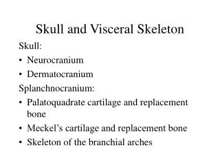
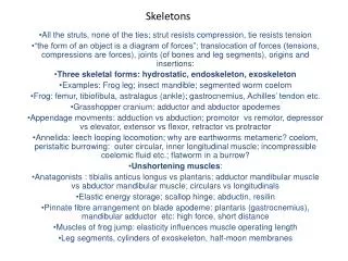
Cranial and Visceral Skeletons. Chapter 9. The cranial skeleton. Three phylogenetic elements: A. Neurocranium (= chondrocranium) Covered in lab (notes on next few slides) Retained in cartilaginous fishes Embryonic cartilaginous braincase in other vertebrates

E N D
Cranial and Visceral Skeletons Chapter 9
The cranial skeleton • Three phylogenetic elements: • A. Neurocranium (= chondrocranium) • Covered in lab (notes on next few slides) • Retained in cartilaginous fishes • Embryonic cartilaginous braincase in other vertebrates • B. Dermatocranium. Dermal bones • C.Splanchnocranium (= visceral skeleton). • Supports gills (gill arches), • In gnathostomes: • jaws • ear • hyoid apparatus
Neurocranium • 1 - protects the brain • 2 - begins as cartilage that is partly or entirely replaced by bone (except in cartilaginous fishes) • Cartilaginous stage: • neurocranium begins as pair of parachordal & prechordal cartilages below the brain • parachordal cartilages expand & join; along with the notochord from the basal plate • prechordal cartilages expand & join to form an ethmoid plate • Cartilage also appears in the • olfactory capsule (partially surrounding the olfactory epithelium) • Otic capsule (surrounds inner ear & also develops into sclera of the eyeball)
Neurocranium (continued) • Completion of floor, walls, & roof: • Ethmoid plate - fuses with olfactory capsules • Basal plate - fuses with otic capsules • Further development of cartilaginous neurocranium = development of cartilaginous walls (sides of braincase) &, in cartilaginous fishes, a cartilaginous roof over the brain • Cartilaginous fishes - retain cartilaginous neurocranium (“chondrocranium”) throughout life
Ventral view of developing chondrocranium in Lacerta Hypophyseal fenestra – allows developmental in(e)vagination of glandular adenohypophysis (anterior pituitary)
Developing Embryo Infundibulum of brain Brain Roof of oral cavity Oral Cavity Rathke’s Pouch Roof of mouth Hypophyseal fenestra
Bony fishes, lungfishes, & most ganoids - retain highly cartilaginous neurocranium that is covered by membrane bone • Cyclostomes - the several cartilaginous components of the embryonic neurocranium remain in adults as more or less independent cartilages • Other bony vertebrates - embryonic cartilaginous neurocranium is largely replaced by replacement bone (the process of endochondral ossification occurs almost simultaneously at several ossification centers)
Neurocranial ossification centers • cartilage surrounding the foramen magnum may be replaced by as many as four bones: • basioccipital (1 element) • exoccipital (2) • supraoccipital (1) • Mammals - all 4 occipital elements typically fuse to form a single occipital bone • Tetrapods - neurocranium articulates with the 1st vertebra via 1 (reptiles and birds) or 2 (amphibians and mammals) occipital condyles) Ethmoid Presphenoid Alisphenoid Basisphenoid Otic Basioccipital Exoccipital Supraoccipital
Occipital condyles • Compare on www.digimorph.org • bird • lizard • Mammal • amphibian What evolved here with respect to the exoccipital elements?
Spinytail lizard badger Caecilian frigatebird
Sphenoid centers form: • basisphenoid bone (anterior to basioccipital) • presphenoid bone • side walls above basisphenoid & presphenoid form: • orbitosphenoid • pleurosphenoid • alisphenoid
Ethmoid centers tend to remain cartilaginous & form • anterior to sphenoid • cribiform plate of ethmoid & several conchae (or ethmoturbinal bones) The ethmoid region is clearly visible within the bisected skull above. In most mammals, the nasal chamber is large & filled with ridges from the ethmoid bones called the turbinals or ethmoturbinals. These bones are covered with olfactory epithelium in life and serve to increase the surface area for olfaction (i.e., a more acute sense of smell). Another ethmoid bone, the cribiform plate, separates the nasal chamber from the brain cavity within the skull.
Otic centers • Otic centers - the cartilaginous otic capsule is replaced in lower vertebrates by several bones: • prootic • opisthotic • epiotic • One or more of these may unite with adjacent replacement or membrane bones: • Frogs & most reptiles - opisthotics fuse with exoccipitals • Birds & mammals - prootic, opisthotic, & epiotic unite to form a single petrosal bone; the petrosal, in turn, sometimes fuses with the squamosal to form the temporal bone
Dermatocranium • Membrane bones of the skull: • Roofing bones (examples on board) • Marginal bones (in upper jaw) • 1° Palate bones • Opercular bones
Basic pattern of "roofing bones" • Crossopterygians – • Series along mid-dorsal line of skull. • paired bones • unpaired bones • Labyrinthodonts – extinct amphibian clade • unpaired bones lost. • series of paired bones resulted • (nasals, frontals, parietals, & dermoccipitals) • Interesting note • Fontanels in teleosts and tetrapods • neurocranium is incomplete dorsally • membranes under the skin ossifying - will eventually close
Marginal bones • Bones of the upper jaw • Pterygoquadrate (=palatoquadrate) cartilage • Vertebrate embryology • 1st upper jaw in development • Cartilaginous fishes - palatoquadrate is the only upper jaw that develops (picture in next slide) • Bony vertebrates - the palatoquadrate becomes covered with dermal bones (premaxillae & maxillae) that make up the adult upper jaw
Placoderm Primitive Cartilage Fish Derived Cartilage Fish Teleosts Ratfish, Lungfish, Tetrapods
Primary palate bones • Palatal bones - • Bony vertebrates - membrane bones form • Fishes and amphibians • “Primary palate” – Common bone on which brain rests and roof of mouth Sharks - cartilaginous • Birds, mammals, & some reptiles • secondary palate (“false palate”) • horizontal partition separating oral cavity into nasal & oral passages. • formed from processes of the premaxillae, maxillae, and palatines.
Opercular bones • Operculum = fold of the hyoid arch that extends back over the gill slits in holocephalans & bony fishes • Tetrapods - no vestiges of opercular bones remain