Download
1 / 88
880 likes | 1.19k Views
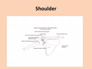
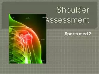
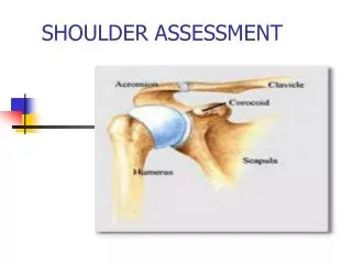
SHOULDER ASSESSMENT. BONY PALPATIONS. Shoulder Complex. Scapulothoracic Articulation. Not a true anatomical joint Resting position of scapula is: Superior angle is level with spinous process T2 Inferior angle level with spinous process T7

E N D
Scapulothoracic Articulation • Not a true anatomical joint • Resting position of scapula is: • Superior angle is level with spinous process T2 • Inferior angle level with spinous process T7 • Medial border of scapula is 5-6 cm or three fingers width from spinous processes
Motions @ Sternoclavicular Joint • SC Joint acts like ball and socket joint • Motions at joint: • Elevation- Depression • Rotation : upward-downward • Rotation: forward-downward; backward-upward
AC Joint Characteristics • Synovial joint • Keeps glenoid fossa continually facing the humeral head • Articular disc between acromion and distal clavicular head • Capsule – lax to allow for complex shoulder motion
AC Joint • Superior and inferior acromioclavicular lig. • Strengthen upper aspect of joint • Limits approx. 90% of anterior-posterior translation
Coracoclavicular Ligament • .: Limits 80% of superior translation of the clavicle- acts as a tie bar to hold clavicle down • Maintains a constant relationship of the scapula on the clavicle
Coracoacromial ligament • Forms roof over humeral head • Prevents upward displacement of humeral head and protects underlying structures • Sharp lateral edge may impinge on bursa and supraspinatus tendon
Superior Transverse Ligament • Bridges lesser scapular notch • Provides a passage for suprascapular nerve
Glenohumeral Joint • Synovial Joint: humeral head articulates with glenoid cavity • Humeral head points medially, backward and tilts upward • Glenoid is ½ as long and 1/3 as wide as the humeral head • Contact area is limited • Surface area of humeral head is 3-4 times larger than the fossa
GLENOHUMERAL AND CORACOHUMERAL LIGAMENTS • Coracohumeral Lig.- One of the most important ligament structures • 1. Blends with rotator cuff, fills space b/t subscapularis and supraspinatus • MaintainsGH relationship • Involved with frozen shoulder
Glenoid labrum • Rim of cartilaginous tissue attached around margin of glenoid fossa • Serves as attachment for ligaments • Deepens articular cavity • Increases glenoid contact with humeral head & serves “chock block” function
Shoulder Bursae • 1. Subacromial or Subdeltoid bursa • 2. B/t coracoid & glenohumeral Jt. Capsule • 3. Summit of the acromion • 4. B/t infraspinatus & joint capsule • 5. B/t teres major & long head biceps • 6. B/t subscapularis & Joint capsule • 7. Tendinous insertion of latissimus dorsi • 8. Behind the coracobrachialis muscle
1st Phase Scapulohumeral Rhythm • Phase I: • Humerus:30 degrees Abduction • Scapula:Minimal movement • Clavicle:0-15 degree elevation
2nd Phase Scapulohumeral Rhythm • Phase II: • Humerus:40 degrees Abduction • Scapula:20 degree rotation • Clavicle:30-36 degree elevation
3rd Phase Scapulohumeral Rhythm • Phase III: • Humerus:60 degrees Abduction • 90 degree lateral rotation • Scapula:30 degree rotation • Clavicle:30-50 degree posterior rotation • up to 30 degree elevation
Biceps tendon mobility • Biceps tendon does not move in the bicipital groove during movement • Humeral head moves over the fixed tendon
Shoulder Patterns: • Closed packed position= 90 degrees abduction and external rotation • Open packed position= arm down by side up to 20-25 degrees abduction
Strength Assessment • Supraspinatus: assessed at 90° of forward flexion in the scapular plane with the thumbs pointed to the floor; downward pressure is resisted by the patient • Test is specific for supraspinatus function, and evaluates cuff strength and integrity
Strength Assessment • External rotators: with the patient’s arm at his side and the elbow flexed to 90°, he will externally rotate as if hitting a tennis ball in a backhanded manner against resistance • Test is specific for the teres minor and infraspinatus muscles
Strength Assessment • Abduction: assessed in the coronal plane against resistance • May be suggestive of either deltoid or cuff deficiency • Subscapularis: with the dorsum of the patient’s hand on his ipsalateral back pocket, instruct him to push backward against resistance
Fractures of Proximal Humeral • Cause: direct blow, a dislocation or the impact received from FOOSH injury • Can be mistaken for shoulder dislocation • Can occur at anatomical neck, tuberosity or surgical neck • Most occur at surgical neck
Humeral Shaft Fractures • Cause: direct blow or Foosh Injury • Type: comminuted or transverse fractures • Signs & Symptoms: Severe pain, swelling, deformity • Complication: radial nerve involvement- loss of wrist and finger extension and sensation over the back of dorsal surface- within 6 months radial nerve should be fine • Treatment: Nonoperative- x-ray views followed by splints and pressure wrap and casting with sling for 1st week
Glenohumeral Dislocations • Most common are anterior displaced with arm abducted and externally rotated • Capsule can remain in tact or be severely damaged as head of humerus in forced out ot glenoid fossa in anterior inferior direction • Secondary labrum injuries – Bankhart Lesion and /or Hill-Sachs Lesions
Anterior Glenohumeral Dislocation • Signs & Symptoms: • Flattened Deltoid contour • Palpation of axilla reveals prominence of humeral head • Athlete carries affected arm in slight abduction and external rotation • Severe Pain with initial dislocation • Tingling and numbness extends down the arm into hand
Hill-Sachs Lesion • Small articular Cartilage defect on the humeral head caused by the impact of humeral head on the glenoid fossa as the humerus dislocates
Recurrent Dislocations and Subluxations • Cause capsule to stretch out allowing for multiple reoccurrences • Athlete complains of arm feeling like it is “Going Dead”- commonly referred to as Dead Arm Syndrome
Tests Used for Shoulder Instability • Sulcus • Aprehension and Relocation
Sulcus • Patient: sitting with arm hanging at the side • Examiner: • grip the arm distal to the elbow • apply a downward force to the humerus while stabilizing the scapula • Positive Test: • a sulcus or indentation appears beneath teh acromion process • Alternate Test: • flex the humerus to 90 and look for a sulcus • this is indicative of inferior instability
Aprehension Test (Anterior) • Patient: Supine with the glenohumeral joint anducted to 90 and the elbow flexed to 90 • Examiner: • support the humerus at midshaft and grasp the forearm proximal to the wrist • passively externally rotate the GH joint by applying pressure to the anterior forearm • Positive Test: • patient displays apprehension that the shoulder might dislocate and resists further movement. • Pain in the anterior capsule of the GH joint • If test is positive then perform the relocation test
Relocation Test • Patient: Supine with the GH joint abducted to 90 and the elbow flexed to 90. • Examiner: • grasp the forearm proximal to the wrist and hold the opposite hand over the humeral head • apply a posterior force to the head of the humerus and maintain it while externally rotating the humerus. • Positive Test: • decreased pain or increased ROM compared to the anterior relocation test
Posterior Apprehension • Patient: supine with the shoulder flexed to 90 and the elbow flexed to 90. The GH joint being tested hangs off of the side of the table • Examiner: • with one hand grasp the forearm, with the other hand stabilize the posterior scapula • apply a downward force to the humerus • Positive Test: • patient displays apprehension and produces muscle guarding to prevent shoulder form subluxating posteriorly