Download

1 / 19
190 likes | 561 Views
Cardiomyopathy and Congestive Heart Failure. NPN 200 Medical Surgical I. Cardiomyopathy . Disease of the heart muscle Cause is unknown Occurs in only 10-20 per 100,000 Results in 30,000 deaths/year 3 types Dilated – both ventricles Hypertrophic – usually die by age 40

E N D
Cardiomyopathy and Congestive Heart Failure NPN 200 Medical Surgical I
Cardiomyopathy • Disease of the heart muscle • Cause is unknown • Occurs in only 10-20 per 100,000 • Results in 30,000 deaths/year • 3 types • Dilated – both ventricles • Hypertrophic – usually die by age 40 • Restrictive – rarest
Cardiomyopathy • Characterized by left and right ventricular failure • Some may be asymptomatic for years and others have acute onset • Stroke volume and cardiac output are decreased • Atypical chest pain which occurs at rest • Progressive and chronic disease
Cardiomyopathy, cont. • Signs and symptoms are dependent upon the type • Dilated • Dyspnea • Fatigue • Left sided heart failure • Cardiomyopathy • Mitral regurgitation (S1 and S2 sounds heard) • Hypertropic • Syncope • Ankle edema • Orthopnea • Angina • Restrictive • Exercise intolerance • Dyspnea • Fatigue • Right sided heart failure • S3 and S4
Cardiomyopathy, cont. • Diagnosis • Echo - primary • Angiography • Radionuclide imaging • Dysrhythmias • Decreased CO with restrictive
Cardiomyopathy, cont • Interventions • Drugs • Diuretics, vasodilators, cardiac glycosides, beta blockers, anticoagulants • Surgery • Excision of the hypertrophied muscle • Mitral valve replacement • Cardiomyoplasty – chest muscle wrapped around the heart • Heart transplant
Nursing Care • Assess • Dyspnea • Cough • Edema • Dysrhythmias • Decreased CO • Need lots of family support and teaching about the disease
Heart Transplant • Transplanted form a donor with comparable weight and ABO compatibility into a recipient less than 6 hours after procurement • Donor must be free of infection, no chest trauma and be declared brain dead, and no malignancies • Most of the cases of transplant are to patients with cardiomyopathy • Patients with a history of noncompliance, depression or inability to cope with stress are not considered good candidates
Heart Transplant, cont. • Recipient is prepared for Open Heart Surgery and placed on cardiopulmonary bypass and the anterior portions of the patients heart are removed and replaced with the donor heart • Post op care is similar to CABG patients • Must be protected from infection by isolation • Must receive immunosuppressant drugs for life, as well as steroids (Solu-Medrol, CellCept, Prograf, Imuran, Sandimmune) • Watch for rejection – temp, malaise, fatigue, dysrhythmias • Monitored by endocardial biopsies • Complications include • Hypertension, ^ cholesterol, obesity, and malignancies
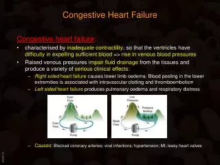
Congestive Heart Failure/Left Sided Heart Failure • Causes the most hospitalizations in patients over the age of 65 • 5 million people on the US are living with heart failure • Inadequacy of the heart to pump blood throughout the body effectively • This deficit causes insufficient perfusion of body tissues with nutrients and oxygen • Causes of heart failure • Coronary artery disease • Acute MI • Cardiomyopathy • Hypertension • COPD • Anemia • Fluid volume overload • Disease of the heart valves
CHF, cont. • 2 ventricles pump independently • Can have right or left sided failure • Usually the left side fails 1st and progresses to failure of both ventricles • May be acute or chronic • May be mild or severe • May be systolic or diastolic failure • May cause pulmonary edema or enlarged liver • Causes retention of sodium and water by the kidneys
CHF, cont. • Right sided failure • May be caused by left ventricle failure, RV infarct, or Pulmonary hypertension • Right ventricle is unable to empty completely • Increased volume and pressure develops in the systemic veins and systemic vascular congestion develops with peripheral edema • Patient may gain fluid weight and have nausea/anorexia, ascites may develop • High output failure • Caused by increased metabolic needs • Septicemia, anemia, and hyperthyroidism
CHF, cont. • Compensation – how the body responds to maintain adequate cardiac output • Sympathetic • Renal • Ventricular hypertrophy
CHF, cont. • Diagnostic tests • History and physical • Chest x-ray shows cardiomegaly with hazy lung fields • Echocardiogram will show enlarged heart and poor contraction of ventricles • BUN and creatinine ^ • Na and Hct may be decreased due to dilution • SAO2 may be decreased • LFT’s may be elevated • B-type Natriuretic peptide (BNP) – produced and released by the ventricles increases
Objective symptoms Left sided failure Anxious Pale Tachycardia Dyspnea, with crackles, wheezes Orthopnea Non-productive cough Later productive cough with frothy, bloody sputum Oliguria Objective symptoms Right sided failure Weight gain Pitting, dependent edema JVD Ascites Decreased UOP Distended neck veins N/V, anorexia CHF, cont.
Nursing Assessment • Vital signs with both apical and radial pulse • HOB elevated • Peripheral pulses • JVD • CVP • Orientation with GCS • Assess for crackles and wheezes • Dependent edema • Weight • Accurate I/O • Abdominal girth • Assess for client and family emotional status
CHF, cont. • Medical treatment • Treat the cause – hypertension, rhythm problems, valve repair • Drugs – cardiac glycosides, diuretics, inotropic agents, vasodilators, ACE inhibitors, beta blockers, Natrecor • Diet – restrictions of sodium and increase of K if diuresis is occuring • Restriciton of H2O • Surgery • Cardiomyoplasty • Heart transplant • Heart reduction surgery
Nursing Interventions • Client education for home care • Must adjust lifestyle • May need O2 • S/S to report to provider • Weight control – contact physician if more than 2 lb weight gain in a week • Dietary management • Medication review • Exercise regimen • Need to work with client to balance activity and rest periods • Monitor for complications • Many have outpatient CHF clinics