Objective
Comparison of Modified ETDRS and Mild Macular Grid Laser Photocoagulation Strategies for Diabetic Macular Edema Sponsored by the National Eye Institute, National Institutes of Health, U.S. Department of Health and Human Services. Compare two laser photocoagulation techniques:


Objective
E N D
Presentation Transcript
Comparison of Modified ETDRS and Mild Macular Grid Laser Photocoagulation Strategies for Diabetic Macular Edema Sponsored by the National Eye Institute, National Institutes of Health, U.S. Department of Health and Human Services
Compare two laser photocoagulation techniques: Modified ETDRS focal photocoagulation (mETDRS) to areas of edema Direct treatment to microaneurysms Grid to diffuse leakage Mild macular grid (MMG) laser technique small mild burns throughout macula in areas with and without edema no direct treatment of microaneurysms Objective
Background • Presumed mechanism of focal photocoagulation include • Closure of microaneurysms • Reduced blood flow leading to auto-regulation and reduced edema • Improved oxygenation leading to auto-regulation and reduced edema • Stimulation of biochemical processes in RPE • Would light widespread laser (mild macular grid, MMG) to the macula be effective? • Pilot clinical trial
Study Design Randomized Clinical Trial (Pilot Study) • Major Eligibility Criteria Assessed: >18 years old Type 1 or type 2 diabetes • Study eye meets the following criteria (subjects allowed 2 study eyes): • Best corrected electronic ETDRS visual acuity score of ≥19 • Definite retinal thickening on clinical exam due to previously untreated DME • Retinal thickness measured on OCT of 250 μm or more in the central subfield or 300 μm or more in at least 1 of the 4 inner subfields • Had no prior laser or other treatment for DME. • Subjects with 2 study eyes: 1 eye was randomly assigned to receive 1 treatment and 1 eye was assigned to receive the other. mETDRS N=162 Eyes MMG N=161
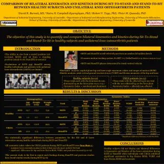
Mild Macular Grid 40 minutes post laser treatment 6 weeks post laser treatment
Follow-up and Treatment Schedule • Measurements by certified Evaluator • Best-corrected electronic ETDRS visual acuity • OCT-measured retinal thickness • Macular laser photocoagulation was repeated if DME persisted and such treatment was warranted in the opinion of the investigator, according to the treatment guidelines. 3.5 Month ± 2 Weeks . . . 8 Month ± 4 Weeks . . . Primary outcome: Change in OCT Secondary Outcome: Change in visual acuity (Method: Repeated measures least squares regression models) 12 Month ± 4 Weeks
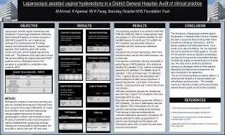
Percent Edema Resolved for Central Subfield Thickness P=0.56 P=0.23 P=0.29
Percent of Edema Resolved for Weighted Inner Zone Thickness P=0.07 P=0.02 P=0.57 Note: Weighted inner zone thickness is a weighted average of the thickness in the central and 4 inner subfields
Percent of Edema Resolved for Maximum Retinal Thickening P=0.93 P=0.26 P=0.57 Note: Maximum thickening is the maximum amount of excess thickness of the central and 4 inner subfields
Percent of Edema Resolved for Retinal Volume P=0.01 P=0.12 P=0.31 Note: Retinal volume is a weighted average of the thickness in the central, 4 inner and 4 outer subfields converted to mm3
Summary • Maximum retinal thickening in the inner zone (within 1500 microns of macular center), central subfield thickness, weighted inner zone thickness and retinal volume decreased in both groups • MMG less effective than mETDRS in reducing retinal thickening • Visual acuity similar in both groups