Download
1 / 14
140 likes | 240 Views
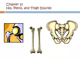
Periacetabular Osteotomy : Intra-articular Work Department of Orthopaedic surgery, University of Toronto, Toronto, Ontario, Canada, . Hip and Pelvis Clinic , C honbuk University Hospital , Jeonju, Korea . E-mail : hugo999@naver.com. Background. Periacetabular Osteotomy

E N D
Periacetabular Osteotomy: Intra-articular WorkDepartment of Orthopaedic surgery, University of Toronto, Toronto, Ontario, Canada, Hip and Pelvis Clinic , Chonbuk University Hospital , Jeonju, Korea . E-mail: hugo999@naver.com
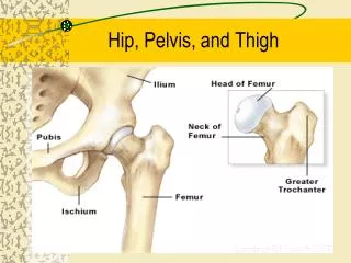
Background • Periacetabular Osteotomy - Goal : Improvement of hip biomechanics by reorienting the acetabulum → postpone degenerative progression Desire of improvement Potential for overcorrection
Background Overlooked intra-articular hip inspection (early experience) Overcoverage of femoral head → iatrogenic FAI Overlooked chondrolabral injury (dysplasia, FAI)
Intra-articular WorkLabral pathology The acetabular labrum - a triangular structure with a basilar attachment to the osseous acetabular rim - a capsular insertion along the external surface - a free intra articular apical margin • Labral injury alone could generate sufficient pain to require intervention • The positive outcome- pain relief after debridement and/or fixation of labral tears,
Intra-articular WorkLabral pathology Labral repair with suture anchor : TOC for unstable hypertrophied labrum Anterior hip arthrotomy Modified Smith-Petersen approach used for Bernese PAO Indirect head of rectus femoris is tagged and mobilized for improved view Capsular incision along the long axis of the femoral neck → AP direction at the level of acetabular rim Simple debridement Labral repair using suture anchor technique is used for labral detachment at the extra-articular osseous insertion Labral detachment → Preservation of the blood supply
Intra-articular WorkLabral pathology Labral takedown and Rim resection Labral refixation with suture anchors fixed to new acetabular rim Correction achieved
Intra-articular WorkChondral pathology • Chondral lesion - Location : anterior and superolateral aspect of acetabulum - Size : 171.7 mm2 • Chondral lesion • Chondromalacic, cleavage, or debonding injury
Intra-articular WorkChondral pathology • The natural history of these lesion and whether chondral lesions are independent source of pain have not been determined. • Nascent chondral lesions identified at the time of PAO represent an opportunity to alter the degenerative cascade.
Intra-articular WorkChondral pathology • In contrast with labral pathology, preOP diagnosis of chondral injury has implications for preOP planning - Hip MR Arthrography : mainstay of soft-tissue hip diagnostic imaging (Specificity ↑, Sensitivity ↓)
Intra-articular WorkChondral pathology Inspection : Patient’s specific hip pathophysiology Diagnostic imaging 2. Rim resection Amount of rim resection to properly address the chondral lesion Staged surgical dislocation → PAO Obtain consent for a possible simultaneous PAO
Femoral head-neck offset • More than 90% of patients treated with PAO require some degree of femoral head-neck offset correction → Intraoperative C-arm fluoroscopy (False profile view, Dynamic impingement view) Assess Intended acetabular correction Femoral head-neck offset Potential exacerbation of FAI
Femoral head-neck offset Cam lesion on femoral head-neck junction burr allows adequate restoration of the femoral head-neck offset Adequate offset achieved