Download
1 / 50
500 likes | 522 Views
Learn about the heart's tissue layers, pericardium structure, myocardium features, and blood supply. Understand the cardiac cycle, conduction system, and cardiac output calculation.

E N D
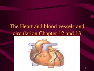
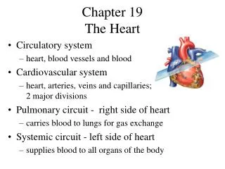
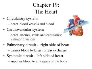
Circulation and the Heart • The circulatory system is a continuous one-way circuit of blood vessels, through which blood is pumped by the heart.
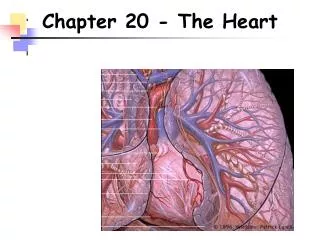
Location of the Heart • Between the lungs • Left of the midline of the body • In mediastinum • Apex pointed toward left
Figure 13-1 The heart in position in the thorax (anterior view). Why is the left lung smaller than the right lung?
Learning Outcomes Describe the three tissue layers of the heart wall. Describe the location and structure of the pericardium and cite its functions. Structure of the Heart
Structure of the Heart Tissue Layers of the Heart Wall • Endocardium lines heart’s interior • Myocardium is thickest layer; the heart muscle • Epicardium is thin outermost layer
Figure 13-2 Layers of the heart wall and pericardium. Which layer of the heart wall is the thickest?
Structure of the Heart The Pericardium • The sac that encloses the heart • Outer fibrous pericardium holds heart in place • Serous pericardium • Parietal layer fused to fibrous pericardium • Visceral layer (epicardium) fused to myocardium • Pericardial cavity is the pace between serous layers
Structure of the Heart Special Features of the Myocardium • Composed of cardiac muscle tissue • Are lightly striated (striped) • Have single nucleus cells • Are controlled involuntarily • Have intercalated disks • Have branching muscle fibers
Figure 13-3 Cardiac muscle tissue viewed under the microscope (x540).
✓ Studies of Disease Checkpoints 13-1 What are the names of the innermost, middle, and outermost layers of the heart wall? 13-2 What is the name of the sac that encloses the heart?
Learning Outcomes Compare the functions of the right and left chambers of the heart. Name the valves at the entrance and exit of each ventricle and identify the function of each. Briefly describe blood circulation through the myocardium. Structure of the Heart
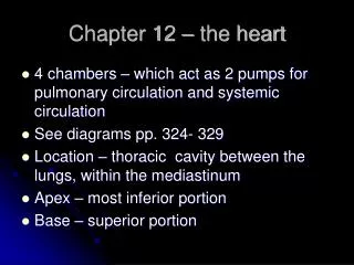
Structure of the Heart Divisions of the Heart • Double pump • Right side pumps blood low in oxygen to the lungs via pulmonary circuit • Left side pumps oxygenated blood to remainder of bodyvia systemic circuit
Structure of the Heart Four Chambers • Right atrium • Receives low-oxygen blood returning from body tissue through superior vena cava and inferior vena cava • Left atrium • Receives high-oxygen blood from lungs • Right ventricle • Pumps blood from right atrium to lungs • Left ventricle • Pumps oxygenated blood to body
Figure 13-4 The heart and great vessels. Which heart chamber has the thickest wall?
Structure of the Heart Four Valves • Atrioventricular valves • Entrance valves • Right atrioventricular (AV) valve (tricuspid valve) • Left atrioventricular (AV) valve (bicuspid valve) • Semilunar valves • Exit valves • Pulmonary valve • Aortic valve
Figure 13-5 Heart valves (superior view from anterior, atria removed). How many cusps does the right AV valve have? The left?
Structure of the Heart Blood Supply to the Myocardium • Coronary arteries • Right coronary artery • Left coronary artery • Cardiac veins
Figure 13-6 Blood vessels that supply the myocardium. What is the largest cardiac vein and where does it lead?
Figure 13-7 Opening of coronary arteries in the aortic valve (anterior view).
✓ Studies of Disease Checkpoints 13-3 What is the heart’s upper receiving chamber oneach side called? What is the lower pumping chamber called? 13-4 What is the purpose of each of the four valves inthe heart? 13-5 What is the name of the system that supplies bloodto the myocardium?
Learning Outcomes Briefly describe the cardiac cycle Name and locate the components of the heart’s conduction system. Explain the effects of the autonomic nervous system on the heart rate. List and define several terms that describe variations in heart rates. Explain what produces each of the two normal heart sounds and identify the usual cause of a murmur. Heart Function
Heart Function Cardiac Cycle • Series of event occurring in the heart during one heartbeat • Systole (active phase, contraction) • Diastole (resting phase)
Figure 13-8 The cardiac cycle. When the ventricles contract, what valves close? What valves open?
Heart Function Cardiac Output (CO) • The volume of blood pumped by the heart per minute • Calculation of cardiac output • Stroke volume (SV): The volume of blood pumped by the heart per heartbeat • Heart rate (HR): The number of heartbeats per minute CO = SV x HR CO = mL/min
Heart Function The Heart’s Conduction System • Produces electrical energy, which stimulates cardiac muscle • Components • Sinoatrial (SA) node (pacemaker) • Internodal pathways • Atrioventricular (AV) node • Atrioventricular bundle (bundle of His) • Purkinje fibers (conduction myofibers)
Figure 13-9 Conduction system of the heart. What parts of the conduction system do the internodal pathways connect?
Heart Function • Although the SA node sets the heart’s pace, the nervous and endocrine systems can influence the heart and allow it to adapt rapidly to changing needs. • Autonomic nervous system (ANS) • Sympathetic nervous system speeds heart up • Parasympathetic system (CNX) slows heart down • Endocrine system • Epinephrine speeds heart up • Thyroxine speeds heart up
Figure 13-10 Autonomic nervous system (ANS) regulation of the heart. Which cranial nerve carries parasympathetic impulses to the heart?
Heart Function Variations in Heart Rates • Bradycardia • Tachycardia • Sinus arrhythmia • Premature beat (extrasystole)
Heart Function Normal and Abnormal Heart Sounds • Normal • Lub • Dup • Abnormal • Organic murmur • Functional
✓ Heart Function Checkpoints 13-6What name is given to the contraction phase of thecardiac cycle? To the relaxation phase? 13-7 What is cardiac output? What two factors determinecardiac output? 13-8 What is the scientific name of the heart’spacemaker? 13-9 What system exerts the main influence on the rateand strength of heart contractions? 13-10 What is a heart murmur?
Learning Outcomes Describe several lifestyle choices that can help maintain heart health. Maintaining Heart Health
Maintaining Heart Health Risk Factors for Heart Disease
Learning Outcome Briefly describe methods used to study the heart. Heart Studies
Heart Studies Methods of Studying the Heart • Stethoscope • Electrocardiograph (ECG or EKG) • Electrodes • Catheterization • Fluoroscope • Coronary angiography • Coronary computed tomography angiography • Echocardiography
Figure 13-11 Normal electrocardiography (ECG) tracing. What is the length of the cardiac cycle shown in this diagram?
Effects of Aging on the Heart Structural Changes • Thinner myocardium • Less flexible valves • Less responsive conduction system Functional Changes • Decreases contraction strength • Decreased cardiac output • Arrhythmia
Case Study Learning Outcome • Referring to the case study, list the emergency and surgical procedures commonly performed following a myocardial infarction and explain why they are done.
Case Study Emergency and Surgical Procedures Commonly Performed Following a Myocardial Infarction • Cardiopulmonary resuscitation • Defibrillation • Administration of thrombolytic medication and nitroglycerine • Administration of morphine • Coronary bypass surgery
Word Anatomy Learning Outcome • Show how word parts are used to build words related to the heart.