Download

1 / 62
630 likes | 896 Views
Secondaries in bone. Content. Introduction Epidemiology Pathogenesis Clinical features Diagnosis Management Medical management Surgical management. Introduction. Introduction.

E N D
Content • Introduction • Epidemiology • Pathogenesis • Clinical features • Diagnosis • Management • Medical management • Surgical management
Introduction • The skeleton is a common site for metastatic spread from epithelial tumours, so patients may present with symptoms from secondary bone cancer to a number of different specialists. • In terms of numbers of patients, the most common cancers to present with bony secondaries are prostate, breast and lung. • Certain patterns of bony spread are common, and an identification of these can guide the need for further investigations. British Orthopaedic Association. Metastatic bone disease: a guide to good practice. London: British Orthopaedic Association; 2001
Introduction… • Secondary breast cancer for example typically presents with multiple metastases affecting the axial skeleton, with a concentration of metastases in the skull, spine, ribs, pelvis and proximal long bones, whereas single metastases or metastases in distal long bones are relatively rarer. • The pathological features of the original breast tumour can be used to predict the risk of systemic recurrence.
Introduction… • The risk of secondary spread in breast cancer is related to- • the grade of the cancer (grade 3 tumours being more likely to metastasize than grade 1), • the size of the original primary tumour (with larger tumours posing a bigger risk) and • the lymph node status of the axilla at the time of the original surgery (with heavily ‘node-positive’ patients – those with metastatic cancer within the axillary nodes at the time of surgical staging – more likely to relapse systemically than ‘node-negative’ ones)
Introduction… • The abnormal investigations need to be interpreted in light of the patient’s risk of developing metastatic disease. • A patient who has been treated for a small grade 1 breast cancer, particularly a tumour of special type such as a tubular carcinoma, is highly unlikely to ever have a recurrence. • A positive bone scan in such a case is therefore more likely to represent either benign disease or secondary spread from a second, undiagnosed primary cancer. Chow E, Finkelstein JA, Sahgal A, Coleman RE. Metastatic cancer to the bone. In: DeVita VT, Lawrence TS, Rosenberg SA, eds. DeVita, Hellman, and Rosenberg's Cancer: Principles & Practice of Oncology. 9th ed. Philadelphia, Pa: Lippincott Williams & Wilkins; 2011: 2192−2204.
Introduction… • On the other hand, a patient with a ‘positive’ bone scan who has been treated for a grade 3 cancer measuring 50 mm in diameter two years previously and who had all 14 axillary nodes involved by tumour at the time of surgery, is much more likely to have metastatic breast cancer. • Certain pathological types of tumour are less likely to spread to bone and these include ovarian cancer, which tends to cause problems throughout the peritoneum, and primary central nervous system (CNS) tumours, which ‘never’ metastasise outside the CNS.
Epidemiology • Secondary bone tumor most frequently occur in patient over 40 year old • Common site of origin are lung, prostate, breast & liver • Common site of deposit are vertebrae, pelvis, femur & ribs • Clinical presentation is extensive and non specific • Most lesion present with osteolytic pattern • Secondary of unknown origin account for 24%
Epidemiology… • Male : Female 2.12 :1 • Mean age 55.7 years & 81.51% Over 41 years • Primary site :- lung 21.8% prostate 13.1% breast 07.4% liver 06.4% G.I.T 05.7% unknown 24.6%
Epidemiology… • Common sites of deposition: • Spine 47.7 % • Pelvis 18.2 % • Femur 15.4 % • Ribs 12.6 % • Multi 20.5 %
Epidemiology… • Primary tumor diagnose before 2dry 29.7 % • Median metastatic time 319 days • Uncertain time in 70.3 % • Osteolytic pattern 80.7 % • Osteosclerotic pattern in 10.5 % • Mixed pattern in 09.8 %
Pathogenesis • Mechanism of Signal Transduction Mediated by Transforming Growth Factor β (TGF-β). • In the extracellular space TGF-β binds either to the type III TGF-β receptor (RIII), which presents it to the type II receptor (RII), or directly to RII on the cell membrane. • The binding of TGF-β to RII then leads to binding of the type I receptor (RI) to the complex and the phosphorylation of RI.
Pathogenesis… • This phosphorylation activates the RI protein kinase, which then phosphorylates the transcription factor Smad2 or Smad3. • Phosphorylated Smad2 or Smad3 binds to Smad4, the common Smad, and the resulting complex moves from the cytoplasm into the nucleus. • In the nucleus, the Smad complex interacts in a cell-specific manner with various other transcription factors to regulate the transcription of TGF-β-responsive genes and mediate the effects of TGF-β at the cellular level.
Pathogenesis… Blobe G et al. N Engl J Med 2000;342:1350-1358
Pathogenesis… Blobe G et al. N Engl J Med 2000;342:1350-1358
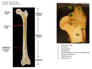
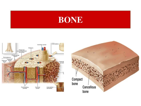
Pathogenesis… • Bone secondaries are common because- • Bone is big organ • Have rich blood supply • Heterogeneous cellular element • Wide orientation along the body • Continues modeling through life • Mode of transmission: • Direct spread • Heamatogenous • Lymphatic
Pathogenesis… • Batson valveless • Connecting inside and outside vertebra and segmentally from pelvis to the skull
Clinical features • Patients typically complain of increasing and persistent pain at the site of bony metastases. • In ‘high-risk’ patients, increasing or new back pain, rib pain or pelvic/hip pain are common symptoms of developing bone metastases. • Rarer symptoms include cranial nerve involvement from base-of-skull metastases, pathological fracture from minimal or no trauma (typically ribs or long bones), sternal pain or sciatica or bladder disturbance from tumour impingement on the sacral formina. Jakofsky DJ et al. Metastatic disease to bone. Hospital physician 2004 Nov: 21-39.
Clinical features… • Vertebral metastases may progress to cause spinal cord compression. • Often the symptoms from this are relatively subtle for a number of weeks, but paradoxically this is the time when early treatment is most effective at preventing catastrophic loss of function. • Therefore any patient at high risk of vertebral metastasis who notices subtle difficulty in walking or a deterioration in leg function, even if no neurological signs are present, should be considered for an urgent magnetic resonance imaging (MRI) scan to exclude spinal cord compression before the condition progresses to paraplegia with loss of sphincter function. • By the time these symptoms occur, treatment is unlikely to restore function.
Clinical features… • Bone metastases may give symptoms of anaemia and investigation is required in more advanced disease. • The full blood count may also show reduced white cell and platelet numbers, owing to bone marrow involvement. • Such haematological abnormalities are particularly common in advanced prostate cancer and these patients may eventually die from pancytopenia and disseminated intravascular coagulation associated with the bony secondaries. Campbells Operative Orthopaedics. Canale ST, Beaty JH editor. 12th edition
Clinical features… • Often the alkaline phosphatase level is raised when bone metastases are present, but the occurrence of hypercalcaemia is less common and often occurs as a paraneoplastic phenomenon in the absence of bone metastases. • It is found more commonly in association with certain cancers, such as lung cancer.
Diagnosis • It is generally unhelpful to check ‘tumour markers’ during investigation of suspected metastatic bone disease, although such tests can be used to assess the efficacy of treatment in patients in whom metastatic disease has been confirmed. • Cancer patients with concerning bone pain should have an initial radiograph of the affected site. • Surprisingly often, this will demonstrate a benign cause for the pain, which requires no further work-up. • Sometimes radiographs will show non-specific abnormality, the nature of which can be clarified by further imaging such as computed tomography (CT) or MRI
Diagnosis… Cancer patient receiving steroids complaining of heel pain. A radiograph demonstrated two calcaneal stress fractures. Cancer patient receiving steroids complaining of hip pain. T1-weighted magnetic resonance image demonstrated right femoral head avascular necrosis
Diagnosis… Cancer patient complaining of left pelvic and hip pain. A radiograph demonstrated acute left pubic rami fractures and left femoral Paget’s disease. Lung cancer patient complaining of generalised limb pain. A knee radiograph demonstrated multifocal periosteal thickening consistent with hypertrophic pulmonary osteoarthropathy (HPOA).
Diagnosis… Patient with haematological malignancy complaining of knee pain. A: A radiograph showed non-specific sclerosis within the distal femur and proximal tibia. B: Subsequent T2-weighted magnetic resonance imaging showed that this was due to medullary bone infarcts.
Diagnosis… • Pelvic insufficiency fractures are a pitfall in the imaging of cancer patients. • A well-recognised cause of bone pain in the elderly, they are especially common in cancer patients who have received steroids or pelvic radiotherapy. • They may occur at several sites in the pelvis but typically the sacral ala and pubic rami. • Often invisible on radiographs, they may be mistaken for metastases on isotope bone scans where they cause intense tracer uptake, or on CT scan where their tendency to heal by sclerosis may be misinterpreted by radiologists primed to search for metastatic disease Coronal computed tomography image from cancer staging scan demonstrating three vertically oriented sacral insufficiency fractures.
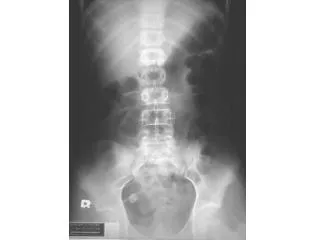
Diagnosis… • If bone pain is due to metastatic disease, a radiograph may show bone destruction (typical of lung cancer), bone sclerosis (typical of prostate cancer), or a mixture of both patterns (common with breast cancer) Radiograph of patient with rectal cancer, showing lytic right inferior pubic ramus metastasis and a colonic stent. Radiograph of patient with prostrate cancer, showing sclerotic metastases in L4, the right sacrum and both acetabula, along with a ureteric stent.
Diagnosis… • Where single long bones such as the femur, tibia or humerus are affected, it is important to assess the risk of pathological fracture through the weakened bone, as elective orthopaedic surgical treatment may be indicated to relieve pain and prevent a future fracture requiring emergency admission Radiograph of patient with breast cancer and myeloma, who sustained a pathological fracture of the midhumeral shaft through a lytic metastasis. Prophylactic intramedullary nailing could have saved this complication.
Diagnosis… • An isotope bone scan is a sensitive method to show metastatic disease at unsuspected and possibly symptomless sites and is commonly used as a staging investigation. • Increased tracer uptake on a bone scan indicates increased activity of osteoblasts, a non-specific reaction to any bony insult, including tumour, trauma, infection, osteoarthritis or inflammatory disease. • This makes bone scans very sensitive to bone pathology but rather non-specific as to its nature. Love C, Tomas MB, Kalapparambath TP et al. Radionuclide bone imaging: an illlustrative review. Radiographics 2003; 23:341–58.
Diagnosis… • When interpreting bone scans, the chance of making a false positive diagnosis of metastatic bone disease is reduced if the pattern of abnormality and underlying likelihood of metastatic disease are taken into account. Asymptomatic cancer patient. A: Isotope bone scan showed increased uptake in sternum and right sacrum, thought suspicious of metastatic disease. B: Subsequent sagittal chest and C: axial pelvic compute tomography (CT) images demonstrated manubriosternal osteoarthritis and a right sacral insufficiency fracture, which account for bone scan findings.
Diagnosis… Breast cancer patient with shoulder pain but low probability of metastatic disease. A: Isotope bone scan showed increased uptake in left humerus, thought suspicious of metastatic disease. B: Subsequent radiograph and C: T1-weighted magnetic resonance images showed a pattern of mineralisation and humeral marrow appearance typical of a benign enchondroma.
Diagnosis… • Computed tomography and MRI scans are also sensitive ways to detect metastatic bone disease but have higher specificity than an isotope bone scan. • In practice, these tests are used to image specific body parts relevant to staging the patient’s cancer and in this situation do not give the comprehensive skeletal coverage offered by a bone scan. Sagittal lumbar spine computed tomography image from lymphoma patient showing sclerotic and lytic bone disease.
Diagnosis… • Vertebral body compression fractures are common in patients with cancer. • Radiographs and bone scans commonly fail to differentiate whether these are due to osteoporosis or metastatic infiltration. • MRI can usually make this distinction, but differentiation between metastasis and a very acute osteoporotic fracture can prove difficult T1-weighted lumbar spine MRI showing multiple chronic osteoporotic vertebral compression fractures characterised by ‘normal’ marrow signal. By contrast, note an acute fracture at D8 exhibiting marrow signal similar to that of metastatic infiltration.
Diagnosis… • In patients with metastases, an MRI scan can uniquely demonstrate the extent of metastatic spinal canal disease. Axial & Sagittal T1-weighted lumbar spine MRI from a lymphoma patient showing multifocal bone marrow infiltration and D10 vertebral body compression fracture.
Management Medical management Surgical management
Medical Management • Adequate analgesia is required and often morphine or related analgesics are necessary. • Non-steroidal antiinflammatory drugs (NSAIDs) are particularly useful in many cases of bony pain, if they can be tolerated. • Systemic treatment of the cancer is often helpful in relieving pain, for example the use of chemotherapy or appropriate hormonal blockade. Oliver TB et al. Diagnosis & Management of bone metastases. J R Coll Physicians Edinb 2011; 41:330–8
Medical Management… • Radiotherapy may also be useful, but there is a limit to how much radiation dose can be given to one area. • Since, extensive irradiation of the axial skeleton has detrimental effects on the synthetic function of the bone marrow and may lead to poorly tolerated chemotherapy (should this be required), due to unexpectedly severe chemotherapy-induced neutropenia caused by the previous radiation treatment. Fairchild A, Barnes E, Ghosh S, et al. International patterns of practice in palliative radiotherapy for painful bone metastases: evidence-based practice? Int J RadiatOncolBiol Phys 2009;75:1281-1628.
Medical Management… • Bisphosphonates have been shown to prevent deterioration in breast cancer and myeloma and, although they have not been proven to be effective in reducing skeletal morbidity in all cancers, are required for the treatment of hypercalcaemia, if present. • All bisphosphonates tend to have some degree of nephrotoxicity, and this needs to be considered when treating patients with abnormal renal function. Mhaskar R, Redzepovic J, Wheatley K et al. Bisphosphonates in multiple myeloma. Cochrane Database Syst Rev 2010; 3: CD003188.
Medical Management… • It is likely that all bisphosphonates have some beneficial activity in this situation; commonly used drugs include zoledronic acid, pamidronate, clodronate and ibandronic acid. • The choice is often determined by patient need and preference (oral versus intravenous) and what trials have been done in the particular tumour site. • If initial analgesic intervention is inadequate, then radiotherapy can be considered.
Surgical Management Orthopaedic surgery: • Since some patients with metastatic bone disease (MBD) will survive three years or more and metastatic pathological fractures rarely unite, it is important to consider fixation of any pathological fracture or lytic lesion. • The decision to operate may be influenced by the histological sub-type of the tumour, as some cancers carry a much poorer prognosis than others, although longevity will not be the only consideration if the operation is expected to provide good palliation. Campbells Operative Orthopaedics. Canale ST, Beaty JH editor. 12th edition
Surgical Management… • Mirel’s scoring system can be used for prophylactic fixation of long bones.
Surgical Management… • Care should be taken to thoroughly investigate bone lesions, especially a solitary lytic lesion (which may not be a metastasis). • There should be no rush to fix the fracture; traction or splintage will keep the patient comfortable while investigations are performed and the case discussed with the lead surgeon for MBD. Janjan N, Lutz S, Bedwinek J, et al. Therapeutic guidelines for the treatment of bone metastasis: A report from the American College of Radiology Appropriateness Criteria Expert Panel on Radiation Oncology. J Palliative Medicine 2009;12:417-426.
Surgical Management… • Surgery aims to relieve pain and restore function and prevent the need for emergency intervention for an unexpected pathological fracture. • The surgery must be planned to allow immediate weight bearing and aim to last the lifetime of the patient. • Surgery for spinal metastases should aim for decompression and stabilisation.
Surgical Management… • Fixation of pathological fractures or lytic lesions tends to have higher failure rates than other types of fracture as the bone rarely heals. • Therefore, for lesions around the hip or proximal femur, a cemented hip prosthesis (standard or tumour prosthesis) is recommended due to its low failure rate. Campbells Operative Orthopaedics. Canale ST, Beaty JH editor. 12th edition