Download
1 / 13
130 likes | 513 Views
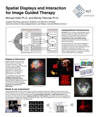
SlicerRT Image-guided radiation therapy research toolkit for 3D Slicer. Csaba Pinter 1 , Andras Lasso 1 , An Wang 2 , David Jaffray 2 , and Gabor Fichtinger 1. 1 Laboratory for Percutaneous Surgery, Queen’s University, Canada 2 University Health Network, Toronto, ON, Canada.

E N D
SlicerRTImage-guided radiation therapy research toolkit for 3D Slicer Csaba Pinter1, Andras Lasso1, An Wang2, David Jaffray2, and Gabor Fichtinger1 1Laboratory for Percutaneous Surgery, Queen’s University, Canada2University Health Network, Toronto, ON, Canada
SparKit project overview • Goal: provide open-source platform for translational clinical research, mainly cancer • Themes: • SlicerRT: radiotherapy toolkit for 3D Slicer • SlicerIGT: Image-guided therapy with 3D Slicer • Funding by Cancer Care Ontario till 2016 • PI & co-PIs: Gabor Fichtinger (Queen’s), David Jaffray (Toronto UHN), Terry Peters (Robarts)
Motivation behind SlicerRT Existing research toolsCERR, PLUNC, dicompyler, etc. Commercial treatment planning systems (TPS) Inconvenient Limitedfeature set Expensive Closed Insufficient user anddeveloper support Open-source? Cover only routineclinical procedures Poor documentation Welldocumented Free Not extensible Extensible Large, non-modularcode base User-friendly Not flexible Flexible Stable Unstable Open-source Platform-independent SlicerRT
Development principles • Leverage existing tools and parallel efforts: 3D Slicer1, Plastimatch2 • “Hub” for RT data analysis and comparison • Cover most common RT research workflows • Free, open-source license (BSD) • Open to integrate algorithms • Extensive documentation XiO® Pinnacle³ Eclipse™ DICOM-RT DICOM-RT DICOM-RT SlicerRT DICOM-RT MATLAB 1S. Pieper, M. Halle, and R. Kikinis, 3D SLICER. Proceedings of the 1st IEEE International Symposium on Biomedical Imaging: From Nano to Macro (Brigham and Women’s Hospital, Boston, MA, 2004), pp. 632–635. 2G. C. Sharp, R. Li, J. Wolfgang, G. Chen, M. Peroni, M. F. Spadea, S. Mori, J. Zhang, J. Shackleford, and N. Kandasamy, “Plastimatch: An open source software suite for radiotherapy image processing,” in Proceedings of the XVIth International Conference on the Use of Computers in Radiotherapy (ICCR) (Amsterdam, the Netherlands, 2010).
General workflow overview SlicerRT components 3D Slicer components
DICOM-RT import • Integrated into core DICOM import plugin mechanism • Data is organized in a smart hierarchy • Supported data types: • RT structure sets→ Contours→ Fiducial point • RT dose map • RT image • RT plan: isocenter, beams • Planning CT, MR, etc. Standard layout after loading phantom dataset
Contour analysis Rasterized volume Ribbon model • Multiple representations(automatic conversion) • Ribbon model • Rasterized volume • Closed surface model • Contour comparison • Dice coefficient • Hausdorff distance • Contour morphology • Expand, shrink • Combine usinglogical operators Closed surface
Dose analysis Dose volume histogram • Dose volume histogram(plot visualization + metrics) • Dose accumulation • Dose comparison (gamma) • Isodose contours / surfaces • Visualize deformation fields • Proton dose computation • Registration • BSplineregistration • Landwarpregistration Isodose contours and surfaces
Example use case Use case: Evaluate the effectiveness of RT plan adaptation • Import, load and visualize data • Register day 1 CT with day 2 CT • Rigid • Bspline • Resample day 2 dose using results • Accumulate doses • Compute and display DVH for all methods • Compare DVH curves and metrics for targetvolume and organs at risk
Software quality • Extensive automatic testing done on multiple platforms every night • Validated against other software packages (Pinnacle, CERR, …) Automatic test infrastructure forautomatic verification and validation Test results reported to the web-based dashboard http://slicer.cdash.org/index.php?project=Slicer4
Extension for 3D Slicer • Collection of RT-specific modules, includes • Distributed as a 3D Slicer extension: can be downloaded, installed, upgraded using the extension manager in Slicer SlicerRT extension in the 3D Slicer app store (free)
Next steps Enhancements planned forthe next 6 months: • DICOM-RT export • Matlab bridge interface:execute Matlab functionsfrom 3D Slicer • Digitally reconstructed radiograph • Support CERR PlanC format • Scripting examples • More testing and validation Current radiotherapy modules (including work-in-progress modules) Detailed plan: https://www.assembla.com/spaces/slicerrt/tickets
Thank you! • Overview paper: Csaba Pinter, Andras Lasso, An Wang, David Jaffray, and Gabor Fichtinger, “SlicerRT: Radiation therapy research toolkit for 3D Slicer”, Med. Phys. 39 (10), October 2012 • Project homepage: http://www.SlicerRT.org/ • Contact: Csaba Pinter (pinter@cs.queensu.ca)