Download
1 / 91
910 likes | 1.08k Views
Nucleic Acids—Big Molecules with a Big Role. Chapter 11. Chapter Outline. 11.1 Components of Nucleic Acids 11.2 Nucleic Acid Formation 11.3 DNA 11.4 RNA and Protein Synthesis 11.5 Putting It Together: The Genetic Code and Protein Synthesis 11.6 Genetic Mutations 11.7 Viruses

E N D
Nucleic Acids—Big Molecules with a Big Role Chapter 11
Chapter 11 Chapter Outline 11.1 Components of Nucleic Acids 11.2 Nucleic Acid Formation 11.3 DNA 11.4 RNA and Protein Synthesis 11.5 Putting It Together: The Genetic Code and Protein Synthesis 11.6 Genetic Mutations 11.7 Viruses 11.8 Recombinant DNA Technology
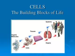
Chapter 11 Introduction • DNA, deoxyribonucleic acid, is the molecule in our cells that stores and directs information responsible for cell growth and reproduction. • DNA is found in the cell’s nucleus. It contains genetic information and is call the genome. • A gene is one part of the genome, and contains information to make a particular protein for the cell.
Chapter 11 Introduction • DNA is long and stringy when isolated and can be seen with the naked eye. • DNA and its counterpart ribonucleic acid, RNA, are made up of repeating building blocks called nucleotides.
Chapter 11 11.1 Components of Nucleic Acids • Nucleic acids are strings of molecules called nucleotides. • Nucleic acids are created from a set of nucleotides in a given sequence. • Each nucleotide has three basic components: • A nitrogenous base • A five-carbon sugar (pentose) • A phosphate functional group
Chapter 11 11.1 Components of Nucleic Acids, Continued • A nucleotide is shown as: Nitrogenous Bases • There are four nitrogenous bases in nucleic acids. Each contains either the purine ring or the pyrimidine ring.
Chapter 11 11.1 Components of Nucleic Acids, Continued • DNA contains two purines, adenine (A) and guanine (G), and two pyrimidines, thymine (T) and cytosine (C). • RNA contains the same bases, except thymine is replaced with Uracil (U).
Chapter 11 11.1 Components of Nucleic Acids, Continued
Chapter 11 11.1 Components of Nucleic Acids, Continued Ribose and Deoxyribose • Nucleotides also contain five-carbon pentose sugars. • RNA contains the pentose ribose (the “R” in RNA) and DNA contains the pentose deoxyribose (the “D” in DNA). • Deoxyribose lacks the oxygen on carbon 2'ofthe pentose.
Chapter 11 11.1 Components of Nucleic Acids, Continued To distinguish between the carbons in the nitrogenous base from those in the sugar rings, a prime symbol (') is added to the carbons in the sugar ring.
Chapter 11 11.1 Components of Nucleic Acids, Continued Condensation of the Components • A pentose and a nitrogenous base can join by a condensation reaction when a nitrogen in the base (N1 of pyrimidines and N9 of purines) bonds to the C1'of the pentose, forming a carbon-to-nitrogen glycosidic bond. • When ribose condenses with the base adenine, a molecule called adenosine is formed.
Chapter 11 11.1 Components of Nucleic Acids, Continued
Chapter 11 11.1 Components of Nucleic Acids, Continued Adenosine is referred to as a nucleoside because it lacks a phosphate group. All nucleosides are formed from condensation of the sugar pentose with a nitrogenous base.
Chapter 11 11.1 Components of Nucleic Acids, Continued • When a hydrogen phosphate (HPO42-) reacts with the –OH on C5' of adenosine, a molecule known as a nucleotide is formed. • The resulting molecule of this condensation is known as adenosine monophosphate, AMP.
Chapter 11 11.1 Components of Nucleic Acids, Continued
Chapter 11 11.1 Components of Nucleic Acids, Continued Naming Nucleotides • Nucleotides contain the name of the nucleoside and the number of phosphates present. An abbreviation is commonly used for the name. • If deoxyribose is found in the nucleotide, a lower case d is inserted at the beginning of the abbreviation.
Chapter 11 11.1 Components of Nucleic Acids, Continued
Chapter 11 11.1 Components of Nucleic Acids, Continued This table summarizes the names of the nucleosides and nucleotides found in RNA and DNA.
Chapter 11 11.2 Nucleic Acid Formation • Many nucleotides linked together form nucleic acids. • Nucleotides are linked together through phosphodiester bonds,where the phosphate oxygens are connected between the 3' and 5' C’s of adjacent sugar molecules. • The formation of a dinucleotide is shown on the next slide.
Chapter 11 11.2 Nucleic Acid Formation, Continued
Chapter 11 11.2 Nucleic Acid Formation, Continued Primary Structure: Nucleic Acid Sequence • A nucleic acid’s primary structure is indicated by its nucleotide sequence. • The backbone of a nucleic acid consists of alternating sugar and phosphate with the bases dangling from the sugar. • Phosphates are connected between the 3' carbon of one sugar and the 5' carbon of a neighboring sugar.
Chapter 11 11.2 Nucleic Acid Formation, Continued • The nucleotide sequence is designated by one-letter abbreviations. • If a single nucleic acid strand is drawn horizontally, the 5' end of the nucleic acid is at the left end, and the 3' end is at the right end.
Chapter 11 11.3 DNA • Nucleic acid sequences, stored as DNA, code for cellular production of protein. • The amount of adenine (A) is always equal to the amount of thymine (T), and the amount of cytosine (C) is always equal to the amount of guanine (G). • The number of purines equals the number of pyrimidines.
Chapter 11 11.3 DNA, Continued Secondary Structure: Complementary Base Pairing • DNA’s secondary structure is described by the interaction of two nucleic acids to form a double helix as proposed by Watson and Crick in 1953. • The double helix is described as a twisted ladder with the sugar–phosphate backbones making the rails and the bases dangling off the backbone, interacting to make the rungs in the center of the ladder.
Chapter 11 11.3 DNA, Continued The two strands are antiparallel to each other, with one strand going in the 5' to 3' direction and the other strand going in the 3' to 5' direction.
Chapter 11 11.3 DNA, Continued • Each rung in the DNA ladder contains one base from each of the strands. The bases interact with each other through hydrogen bonding. • All rungs are of the same length and consist of a purine and a pyrimidine. The pairs A–T and G–C are called complementary base pairs. • Adenine forms two hydrogen bonds to thymine, and guanine forms three hydrogen bonds to cytosine. The DNA in one human cell contains about 3 billion of these base pairs.
Chapter 11 11.3 DNA, Continued
Chapter 11 11.3 DNA, Continued Tertiary Structure: Chromosomes • The compact structure of DNA, caused by the double helix twisting on itself, constitutes its tertiary structure. The further twisting of DNA is called supercoiling. • The 3 billion base pairs of DNA in one human cell would stretch out as a double helix to about 6 feet in length.
Chapter 11 11.3 DNA, Continued • DNA in a human cell is separated into 46 pieces (23 from mother, 23 from father) that are supercoiled around proteins called histones. • DNA pieces twist about histones and are packed into chromosomes. Chromosomes are an efficient package for large amounts of DNA information.
Chapter 11 11.3 DNA, Continued
Chapter 11 11.4 RNA and Protein Synthesis • RNA, like DNA, is a strand of nucleotides, and is responsible for protein synthesis. • There are differences in the nucleotides of RNA and DNA. • RNA does not contain thymine, instead it contains uracil. Uracil is complementary to adenine, and forms two hydrogen bonds with adenine.
Chapter 11 11.4 RNA and Protein Synthesis, Continued RNA Types and Where They Fit In There are three types of RNA found in cells: • Messenger RNA (mRNA) • Ribosomal RNA (rRNA) • Transfer RNA (tRNA) • Messenger RNA and Transcription
Chapter 11 11.4 RNA and Protein Synthesis, Continued Messenger RNA and Transcription • The process of making protein requires two steps. • The first step is called transcription, which is a process of making a gene copy from the DNA. • In transcription, DNA unwinds temporarily and a complementary strand is made from one of the strands. • This complementary copy is called the messenger RNA (mRNA). • mRNA is a single strand of complementary bases of the original DNA, and copying is catalyzed by the enzyme RNA polymerase. • mRNA travels to an organelle called the ribosome, where the mRNA sequence is processed into protein.
Chapter 11 11.4 RNA and Protein Synthesis, Continued Ribosomal RNA and the Ribosome • A ribosome is made up of ribosomal RNA (rRNA)and protein. It is the protein factory in the cell. • Information in mRNA is interpreted into an amino acid sequence in the ribosome.
Chapter 11 11.4 RNA and Protein Synthesis, Continued Ribosomal RNA and the Ribosome, Continued • Ribosomes are composed of two rRNA/protein subunits called the small and large subunit. • mRNA fits in a groove on the small subunit with the bases pointing toward the large subunit.
Chapter 11 11.4 RNA and Protein Synthesis, Continued Transfer RNA and Translation • The second step in protein synthesis occurs in the ribosome and is called translation. • Transfer RNA (tRNA)is the facilitator for this process. • tRNA is T-shaped due to areas of hydrogen bonding by complementary bases. • tRNA has a three-base sequence (triplet) called an anticodon at its anticodon loop. • The anticodon hydrogen bonds to three complementary bases on mRNA.
Chapter 11 11.4 RNA and Protein Synthesis, Continued Transfer RNA and Translation, Continued • tRNA has a place on the opposite end of the anticodon called the acceptor stem,where it can bind an amino acid through esterification. • Each one of the 20 amino acids has one or more tRNAs that can transfer it to the ribosome for incorporation into a growing protein chain.
Chapter 11 11.4 RNA and Protein Synthesis, Continued Transfer RNA and Translation, Continued
Chapter 11 11.5 Putting It Together: The Genetic Code and Protein Synthesis The Genetic Code • A given triplet in mRNA contains a base sequence, transcribed from DNA, that translates to a specific amino acid. This triplet is called a codon. • For example, the sequence UUU in a mRNA specifies the amino acid phenylalanine, GGG specifies glycine, and CGC specifies arginine.
Chapter 11 11.5 Putting It Together: The Genetic Code and Protein Synthesis, Continued • The tripeptide produced from the mRNA codons UUUGGGCGC is Phe–Gly–Arg. • The genetic codeassigns all 20 amino acids to codons of mRNA. There are 64 possible codon combinations from the four bases A, G, C, and U.
Chapter 11 11.5 Putting It Together: The Genetic Code and Protein Synthesis, Continued • The three codons UGA, UAA, and UAG are stop signals for protein synthesis. • The triplet AUG serves two purposes in protein synthesis. • It represents the start codon initiating protein synthesis if it is at the 5' end of an mRNA. • It codes for the amino acid methionine if it is found elsewhere in mRNA.
Chapter 11 11.5 Putting It Together: The Genetic Code and Protein Synthesis, Continued
Chapter 11 11.5 Putting It Together: The Genetic Code and Protein Synthesis, Continued Protein Synthesis In review, protein synthesis involves: • Transcription,during which a complementary copy of DNA, called mRNA, is created by RNA polymerase. mRNA travels out of the nucleus to the ribosome. • tRNA activation, during which atRNA synthetase attaches the correct amino acid to the acceptor stem of the tRNA.
Chapter 11 11.5 Putting It Together: The Genetic Code and Protein Synthesis, Continued
Chapter 11 11.5 Putting It Together: The Genetic Code and Protein Synthesis, Continued In review protein synthesis involves: • Translation: – Protein synthesis starts with the start codon. – Activated tRNA, with methionine attached, enters the ribosome and hydrogen bonds to mRNA. – The second activated tRNA, matching the next codon on mRNA, enters. The two amino acids join. – The first tRNA leaves and the tRNA with the dipeptide attached shifts into the first position, a shifting calledtranslocation. – This process continues over and over until a protein chain emerges.
Chapter 11 11.5 Putting It Together: The Genetic Code and Protein Synthesis, Continued In review protein synthesis involves: • Termination: – Eventually the ribosome encounters a stop codon, and protein synthesis stops. – The protein chain is released. – The initial amino acid methionine is often removed from the beginning of the chain. – The protein chain folds into its tertiary structure that makes it a biologically active protein.
Chapter 11 11.5 Putting It Together: The Genetic Code and Protein Synthesis, Continued
Chapter 11 11.6 Genetic Mutations A change in a DNA nucleotide sequence is called a mutation. What happens to protein synthesis after a mutation? • No change in protein sequence. Only 2.5% of DNA encodes for genes. The rest is “junk” DNA. These types of mutations are called silentmutations. • A change in protein sequence occurs, but it has no effect on protein function. For example, a mutation may occur that results in an amino acid that has similar polarity and size to that of the original amino acid.
Chapter 11 11.6 Genetic Mutations, Continued A change in protein sequence occurs that affects protein function. For example, a mutation could occur that involves replacing a nonpolar amino acid with a polar amino acid. The structure and function of the resulting protein could change. A codon could be mutated to a stop codon. Bases could be inserted or deleted in a DNA strand.
Chapter 11 11.6 Genetic Mutations, Continued Sources of Mutations • Sometimes errors occur at random when DNA replicates itself. This is called a spontaneous mutation. • Mutagens, like environmental agents or radiation can cause mutations. • Sodium nitrite, used as a preservative in processed meats like hot dogs and bologna, is a chemical mutagen.