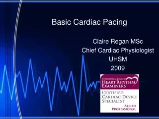
External Cardiac Pacing
External Cardiac Pacing. Dr PG Jones MBChB, FACEM Emergency Medicine Specialist Auckland And Starship Hospitals. History. 1791 Galvani Frog experiments 1892 Duchenne Resuscitated child (submersion ) One leg electrode, tapped chest with other. Zoll. 1950’s

External Cardiac Pacing
E N D
Presentation Transcript
External Cardiac Pacing Dr PG Jones MBChB, FACEM Emergency Medicine Specialist Auckland And Starship Hospitals
History • 1791 Galvani • Frog experiments • 1892 Duchenne • Resuscitated child (submersion ) • One leg electrode, tapped chest with other
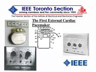
Zoll • 1950’s • 1st successful TCP and monitor • 3cm electrode • 120V AC for 2msec • 1980’s • 80cm2 electrode, 40msec pulse duration • stimulation threshold x 6 • More tolerable = renewed interest • FDA approval 1982
Indications • Symptomatic Bradycardia • 50-100% survival to discharge rates • AMI with certain ECG rhythms • Mobitz type II second-degree AV block • Third-degree heart block • Bilateral BBB • Newly acquired or age-indeterminate LBBB, LBBB and LAFBa, RBBB, and LPFBa • RBBB or LBBB and first-degree AV block
Indications • Asystole ? • Most studies no benefit • Benefit shown for <5min post arrest • 2/5 survived neurologically normal • Tachyarrythmia • 57-95% termination of VT • 4-24% acceleration of VT
How To Do It • Inform the patient • Plug in module • Attach pads • Set rate 70 • Dial up mAmps • Set mode (demand first) • Start • Monitor and adjust as needed
Capture • Feel the pulse • mechanical capture • 2nd monitor to determine electrical capture • Unless monitor blanks out skeletal muscle contraction • Ultrasound
Not Capturing Native Beat Paced Beat IABP
Current • 65-100mA (Unstable patient) • 50-70mA (Volunteer) • 90% tolerate for 15min • Pain Current / area (up to 10cm2)
Energy • 100mA for 20msec = 0.1J (with normal TTR, 50 Ohm) • Threshold for discomfort 1-2J (Skin tingling) • Does not damage the myocardium
Discomfort • Skeletal muscle contraction is the cause • Often the limiting factor • Attempt AP placement to minimise • Left scapula and midline chest • Use lowest effective current • Sedation as needed • CPR is safe!
Haemodynamics • Cardiac arrest and CHB • Comparable to transvenous pacing • Sinus • Reduced cardiac output • No ‘atrial kick’ (atrial capture threshold too high)
References • Bocka JJ eMedicine http://www.emedicine.com/emerg/topic699.htm • Updated 2002 April 24, excellent summary article • Bocka JJ Ann Emerg Med 1989 Dec; 18(12):1280-6 • Hedges JR Pacing Clin Electrophysiol 1991 Oct;14(10):1473-8 • Barthell E Ann Emerg Med 1988 Nov;17(11):1221-6 • Klumbies A Z Gesamte Inn Med 1988 Jul 1;43(13):348-52.