Download
1 / 62
620 likes | 648 Views
This review explores the process of implantation and placentation in rodents, with a focus on the five phases of implantation. It also highlights the role of various factors, such as mucins, integrins, endocannabinoids, and growth factors, in the successful implantation of the blastocyst.

E N D
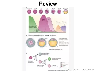
Review Wang and Dey, 2006 Nature Reviews 7:185-199
Lecture X : Comparative Implantation Wang and Dey, 2006 Nature Reviews 7:185-199
Implantation Rodents (Mouse) Contact of the blastocyst with the uterus is the first step in the relationship of a mammal embryo with its environment. This contact leads to implantation and placentation. Implantation can be divided into five separate phases: 1. Hatching Phase - 2. Apposition Phase - 3. Adhesion Phase - 4. Invasion Phase - 5. Postimplantation Phase - Blastocyst loss of Zona pellucida Hatched blastocyst aligns and orients itself in preparation for attachment Attachment between uterine surface epithelium and trophoblast After blastocyst becomes firmly anchored to the uterine surface, the trophoblast cells begin to penetrate the uterine epithelial layer Trophoblast cells cease invasive activity and the maternal-fetal relationship is stablized
Mesometrial Endometrium Uterine epithelium Zona pellucida ICM Trophectoderm Antmesometrial Blastocyst enters uterus on Day 4 Hatching from the Zona Pellucida Compartive Placentation (ed. D.H. Steven), 1975
Note the closure of the lumen There is extracellular material between uterine epithelium and trophectoderm Apposition Phase Attachment Phase Blastocyst comes to lie along the antimesometrial side of uterus. Note that ICM is oriented toward the mesometrial side of uterus. Orientation may be caused by the shape of embryo which is larger at the embryonic pole. Treatment with Relaxin can disrupt the orientation of the blastocysts. Decidualization begins following adhesion in rodents. Surface changes in the uterine epithelium permit adhesion between trophectoderm and uterine surface epithelium Compartive Placentation (ed. D.H. Steven), 1975
Receptivity of Mouse Uterus to Blastocyst Attachment Mucins are large O-linked glycoproteins with ectodomains that extend away from the surface of the cell apical border. Mucins may sterically block access to the cell membrane and function as anti-adhesive molecules. Muc-1 also referred to as epsialin, may function in the time of blastocyst attachment in mammals. Muc-1 is lost from the uterine epithelial apical surface during diestrous in the mouse. It is expressed only on the apical border during the first 3 days of the estrous cycle.
Muc-1 expression Muc-1 antibody (green) Perlecan antibody (red) Basement membrane Lumenal Epithelium Glandular Epithelium Muc-1 immunocytochemical staining A. Proestrous B. Estrous C. Metestrous D. Diestrous Only in glandular epithelium, loss of intensity in surface Western blot of Muc-1 Surveyor et al. 1995, Endocrinology 136:3639
Northern Blot A, B, and C = Days 1, 2 and 3 pregnancy D = Day 4 Note: Loss of Muc-1 is associated with time of implantation. Surveyor et al. 1995, Endocrinology 136:3639
Northern Blot Estrogen stimulates Muc-1 expression. Progesterone inhibits Muc-1 expression. Lane 1 = Day 1 pregnancy Lane 2 = Day 4 pregnancy Day 4 of pregnancy mice ovariectomized and given either P4 (lane 3) or E plus P4 (lane 4). Surveyor et al. 1995, Endocrinology 136:3639
Model for Muc-1 function as an antiadhesion moleucle Surveyor et al. 1995, Endocrinology 136:3639
Role of epithelial surface mucins in steric inhibition of embryo-endometrial interactions Aplin, 1997; Rev. Repod. 2:84-93
Lipid Hydrolysis Prostaglandin Synthesis Increased Membrane Permeability Moulton and Koenig, 1986, In Nidation (ed. Yoshinaga) pp95-109
Heparin Sulfate Proteoglycans av integrin through a bridging ligand Heparan sulfate binding to a basic protein. Also possibly Chondroitin sulfate Cadherin binding Interaction of trophoectoderm and endometrial cells at implantation. Aplin, 1997; Rev. Repod. 2:84-93
Integrins a has 17 subunits b has 8 subunits
Endocannabinoid Signaling Endocannabinoids (Marijuana) target receptors (CB1 and CB2) in the mouse embryo. CB1 localized in trophectoderm and not ICM CB1 in oviduct and uterus Knockout of CB1 causes defects in development and survival High levels of the endocannabinoid, Anandamide (AEA), inhibit early embryo development. However, low levels are needed and stimulate trophoblast development. Anandamide levels are also important for implantation. Implantation competency requires down-regulation of AEA binding to the blastocyst. Coordinated down-regulation of CB1 on blastocyst and decrease in AEA from uterus. Women with elevated peripheral AEA levels have spontaneous pregnancy loss. Sun and Dey. Mol Cell Endo (2008) 286:S1-S11
Role of Growth Factors in Preimplantation Development IL-6 will bind to LIFr as well Colony Stimulating Factor (CSF-1) has been shown to be involved with implantation too! Leukemia Inhibitor Factor has been demonstrated to be involved with implantation in the mouse.
LIF appears following estrogen in delayed implantation LIF stimulates proteinase expression in the uterus. Like - MMPs urokinase type plasminogen activator. LIF not present in delayed implantation
Histological features of the uterus in LIF-null and wild-type mice. Semi-thin resin sections from days 5 and 6 of pregnancy were stained with toluidine blue. The insets show high magnification of differentiated polygonal stromal cells in the wild-type and undifferentiated fibroblast-like stroma cells in the LIF-null mice on day 6 of pregnancy. E = embryo, scale bars = 50 µm (Reprinted from Developmental Biology, vol 281, Fouladi-Nashta et al.
LIF Activation stimulates Heparin Binding Epidermal Growth Factor
Autocrine versus juxtacrine signaling modes. In the simplest model, autocrine signaling is regulated by the removal of the prepro-extension from the membrane-anchored ligand (step 1) followed by its controlled release from the membrane (step 2). Orientation restrictions are responsible for the release requirement. In the case of juxtacrine signaling, prepro-extension release is required (step 1), followed by binding to an auxiliary molecule on a neighboring cell (step 2). Note that autocrine ligands bind to the cell that produced them, and juxtacrine ligands bind to a neighboring cell.
Activation of Implantation by LIF induces HB-EGF in LE of Endometrium Amphiregulin HB-EGF Epiregulin
Immunofluorescence staining for Cox-2 and oncostatin M (OsM) in LIF-null and wild-type mouse uterus on days 5 and 6 of pregnancy. Cox-2 protein is strongly expressed in the LE and underlying stromal cells (arrows) at the implantation site on day 5 and expression extended deeper into the stroma by day 6. In LIF-null mice, expression was limited to the LE cells and only a few stromal cells expressed Cox-2 in the day-6 uterus. The pattern of expression for OsM protein in wild-type mice was similar to Cox-2. In LIF-null animals, OsM was completely absent around the embryo on days 5 and 6 of pregnancy. E = embryo, scale bar = 100µm (Reprinted from Developmental Biology, vol 281, Fouladi-Nashta et al.
Cross et al. 1994 Science 266:1508
At day 4.5 of mouse development, the blastocyst attaches to the uterine epithelium and uterine lumenal closure occurs. A surge of estrogen from ovary stimulates implantation. Estrogen releases the cytokines such as LIF, IL-1, HB-EGF and CSF-1. Adhesion molecules induce binding through lectin- carborhydrate and integrin-integrin interactions Cross et al. 1994 Science 266:1508
Implantation Preimplantation Sherman et al. 1979, In: Matermal Recogniton of Pregnancy, Cibia foundation symposium 64, pp 33-52
Trophoblast cells begin to penetrate epithelium Implantation Chamber Decidual cells form Decidualization - uterine stroma undergoes a transformation, fibroblasts proliferate and enlarge stromal area Invasion Phase The uterine epithelium degenerates (apoptosis) and trophoblast cells come into contact with decidua Postimplantation Phase Trophoblast cells lose their invasiveness Compartive Placentation (ed. D.H. Steven), 1975
Mouse blastocyst implantation Day 7 p.c. Note: Position and orientation of blastocyst. Surface epithelium is intact Decidual Reaction Blastocyst Mouse blastocyst implantation Day 8 p.c. Note: Blastocyst is inside stroma with loss of uterine epithelium Uterine Lumen Perry, 1981; J. Reprod. Fert. 62:321
Tachi and Tachi, 1986, In Nidation (ed. Yoshinaga) pp158-182
Day 5 blastocyst implantation Tung et al. Biol Reprod 35:1045
Tachi and Tachi, 1986, In Nidation (ed. Yoshinaga) pp158-182
Rat Implantation site on Day 7 p.c. Luminal epith Collagen Fibers Decidual cells Tung et al. Biol Reprod 35:1045 Capillary
Blastocyst Implantation Chamber Enders and Schlafke, 1979, In: Matermal Recogniton of Pregnancy, Cibia foundation symposium 64, pp 3-32
Tachi and Tachi, 1986, In Nidation (ed. Yoshinaga) pp158-182
Restriction of dye from the primary decidual zone Tung et al. Biol Reprod 35:1045
Role of Metalloproteinases and Tissue Inhibitor of Metalloproteinases
Urokinase-type plasminogen activator Plasminogen MMPs can be regulated by steroids- P4 negative role Cytokines - IL-1, TNF-a MMP secreted in their latent form and most be cleaved to be activated Tissue inhibitor of metalloproteinases a-macroglobulin Salamonsen, 1999;Rev. Reprod. 4:11-22