Pancytopenia
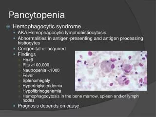
Pancytopenia. Hemophagocytic syndrome AKA Hemophagocytic lymphohistiocytosis Abnormalities in antigen-presenting and antigen processing histiocytes Congenital or acquired Findings Hb <9 Plts <100,000 Neutropenia <1000 Fever Splenomegaly Hypertriglyceridemia Hypofibrinogenemia

Pancytopenia
E N D
Presentation Transcript
Pancytopenia • Hemophagocytic syndrome • AKA Hemophagocyticlymphohistiocytosis • Abnormalities in antigen-presenting and antigen processing histiocytes • Congenital or acquired • Findings • Hb<9 • Plts <100,000 • Neutropenia <1000 • Fever • Splenomegaly • Hypertriglyceridemia • Hypofibrinogenemia • Hemophagocytosis in the bone marrow, spleen and/or lymph nodes • Prognosis depends on cause
At Risk Population • Hereditary cutaneous syndrome • Neurocutaneous syndromes • Chromosomal abnormalities • Hereditary or acquired immunodeficiency • Congenital malformations or syndromes • History of prior cancer • Intrauterine or postnatal exposure to certain agents • Radiation • Metabolic diseases • Identical twin with malignancy • FH of Wilms or RB
Warning Signs • Pallor • Fatigue • Petechiae • Fever w/o source • Bone pain • Weight loss • Anorexia • HA, vomiting, lethargy • Lymphadenopathy • Hepatomegaly • Splenomegaly • Abdominal Mass • Testiculomegaly • Metabolic abnormalities • Hyperkalemia • Hyperuricemia • Hypocalcemia
Lymphadenopathy • Warning signs • Large • Hard • Fixed • Persistent
Mediastinal Mass • Anterior • Lymphomas • Hodgkin and non-Hodgkin • T-cell leukemia • Germ cell tumors • Thymomas • Middle • Lymphomas • LAD secondary to leukemia • Posterior • Neuroblastoma • Ewing sarcoma
Mediastinal Mass • Superior vena cava syndrome • Obstruction of venous return to the heart • Facial swelling • Plethora • Wheezing • Cough • Upper extremity edema
Abdominal Mass • Causes • HSM • Mass of alternative origin • Consequences • Ascites • Superficial venous distention • Look for DVTs • Lower extremity edema • Scrotal/labial swelling • Renal dysfunction • Respiratory difficulty
Leukemia • Acute leukemia • Malignant replacement of marrow • Most common cause of cancer in children • Blast cells • Clonal expansion of a cell at a specific stage of lymphoid or myeloid development
Leukemia • Signs • Fever • Fatigue • Pallor • Organomegaly • Lymphadenopathy • Gingival hyperplasia • Testicular enlargement • Hemihypertrophy
Leukemia • Signs • Bone • Pain • Metaphyseallucencies • Growth arrest lines • Osteopenia • Lytic bone lesions • ALL 75% • 2-5y • AML 20% • Older children • Worse prognosis
Acute Lymphocytic Leukemia (ALL) • Most common pediatric malignancy • Poor prognostic signs • <2y • WBC>50 • T-Cell • Good prognostic sign • Pre-B cell
Hodgkin’s Lymphoma • Presentation • Teens • Non-tender enlarged cervical lymph nodes • Weight loss • Fevers • Night sweats • Slow-growing nodal progression • Cervical • Mediastinal • Abdominal • Pelvis • Finding • Reed Sternberg cell on biopsy
Non-Hodgkin Lymphoma • Peak incidence • 5-8y (younger than HL) • Presentation • Palpable mass • Usually head and neck • 25-50% present with abdominal involvement • Mediastinal mass • Pleural effusions • Abdominal pain • Testicular enlargement
Question 9 The mother of a 6 month old child arrives for a sick visit. Mom says that the patient has vomited his morning bottle almost every day for the last month. She attributed this symptom to reflux since he has also been fussy in the mornings, but this morning his eyes look funny too (picture). Of the following, what is the most likely diagnosis? • Retinoblastoma • Neuroblastoma • Langerhans cell histiocytosis • CNS tumor • Lymphoma
CNS Tumors • Second most common malignancy in children • Signs of increased ICP • Morning HA • With vomiting • Papilledema • Neurologic deficits • Cranial nerves • Macrocephaly • Bulging fontanelle • AMS
CNS Tumors • Brain Tumors • Most common type of solid tumors • Most are infratentorial • Cerebellum • May cause ataxia • Spinal cord tumors • May present anywhere • Signs • Pain on percussion over vertebral column • Weakness of affected muscle group • Bowel-bladder dysfunction • Parestheisias • Changes in gait
Brain Tumors • Medulloblastoma • Poorly differentiated neuroepithelial cells • Cerebellar • Seeding of subarachnoid space may occur • Craniopharyngioma • Supratentorial (thalamus or hypothalamus) • Signs • FTT • Abnormalities in sexual development • Visual field distubances
Brain Tumors • Astrocytoma • Supra or infratentorial • Signs depend on location • Ependymoma • Supra or infratentorial • 4th ventricle is most common • Obstructive hydrocephalus is most common presenting condition • Brainstem glioma • Insidious symptoms • Isolated cranial nerve deficits • Long tract signs
Retinoblastoma • Leukocoria with an absent red reflex (60%) • Strabismus may also be present (20%) • Genetic predisposition • If parent had it in both eyes • 50% risk for child • If parent or sibling had it in one eye • 5% risk for child • Only 5% have a FH • At risk for osteosarcoma later in life
Neuroblastoma • Most common extracranial solid malignancy of childhood • Most present • <5y • Extensive locoregional disease (3) • Widespread metastasis (4) • Embryonic tract of neural crest • Neck to pelvis • >50% adrenal or juxtarenal
Neuroblastoma • Characteristics • Multilobular • Firm • Retroperitoneal • Encase vessels • Cross midline
Neuroblastoma • Signs • Abdominal distention/mass • FTT, weight loss, anorexia • UTI from obstruction • Horner’s syndrome • Paralysis and/or weakness • Invasion of spinal cord or peripheral nerves • Cerebellar ataxia
Neuroblastoma • Signs • Nodular skin lesions • Racoon Eyes • Metastatic disease to the orbit • Ecchymoses in the periorbital area
Neuroblastoma • Signs • Heterochromia • Although usually benign may be associated with cervicothoracicneuroblastoma
Neuroblastoma • Paraneoplastic • Opsoclonus-myoclonus syndrome • Random eye movements and myoclonic jerking • More favorable prognosis • Kerner-Morrison syndrome • Intractable secretory diarrhea • Hypokalemia • Dehydration • Due to secretion of vasoactive intestinal peptide • Resolves with tumor resolution • Catecholamines can also result in irritability and HTN
Neuroblastoma • Lab eval • Urine • Vanillyl-mandelic acid • Homovanillic acid • Ferritin • Neuron-specific enolase • LDH • Biopsy • Radiology • CT • Bone scan • MIBG
Neuroblastoma • Radiology • Stippled calcifications • Retroperitoneal mass that crosses midline • Intimate involvement of vascular structures • Aorta • Vena cava • Renal vessels • Mesenteric artery • IVP • Drooping lily
Neuroblastoma • Poor prognosis • <25% survival • Variant • Stage IV-S • Extensive local and widespread metastases to bone, skin and liver • Children <1y • Spontaneous resolution
Wilms Tumor • Second most common abdominal malignancy in childhood • Most common renal tumor of childhood • Can present • <5y (median age 3.5y) • Bulky abdominal mass • Hematuria • HTN • Severe pain secondary to bleeding or rupture • Most children are asymptomatic
Wilms Tumor • PE • Large bulky tumor • Subcostal • Firm • Smooth • Immobile • Nontender • Does not cross midline • Aniridia • WAGR syndrome • Wilms, aniridia, genital abnormalities, MR • Hemihyptertrophy
Wilms Tumor • Radiology • CT chest and abdomen • Site of origin • Bilateral disease • Extent of local disease • Involvement of vessels • Renal veins • IVC • RA
Wilms Tumor • Echo • Intracaval thrombus • RA thrombus • Chemo before resection • Bone scan • Mets • Lungs • Lymph nodes • Liver • Bone
Hepatic Neoplasms • Rare in childhood • 2 most common • Hepatoblastoma • Young children • >75% less than 3y • May present with hemihypertrophy • Hepatocellular carcinoma • Older children • Elevated α-fetoprotein • Often more present in hepatoblastoma • Nonspecific signs
Rhabdomyosarcoma • Most common soft tissue sarcoma of childhood • Histology • Striated skeletal muscle • Arise anywhere in the body • Prognosis depends on site and histology
Rhabdomyosarcoma • 2 peak incidences • 2-6y • 15-19y • Younger patients • Head • Neck • GU • Adolescents • Extremity • Truncal • Paratesticular
Rhabdomyosarcoma • Orbital • Proptosis • Macrocephaly • Increased ICP • Head and Neck • Otorrhea • Intraoral extension • Testicular
Rhabdomyosarcoma • Sarcoma botryoides • Grape-like morphology • May involve • Vagina (classic) • Bladder • Nasopharynx
Teratomas • Signs • Apparent on PE • Most commonly in sacrococcygeal area • May appear • Pineal • Mediastinum • Retroperitoneum • Ovary/Testes • Embryonal • Contain tissue from all 3 germ cell layers • 20% malignant
Question 10 A 16 yo WM presents to your clinic with the complaint of fever and pain in his leg. The patient recalls a minor injury (contusion to the area) during football practice 2 weeks ago but his symptoms resolved prior to this presentation. An X-ray is taken and shown. What is your diagnosis? • Osteomyelitis • Ewing Sarcoma • Osteosarcoma • Leukemia • Rhabdomyosarcoma
Primary Bone Tumors • Adolescence • Signs • Persistent pain • Osteosarcoma • Development coincides with linear bone growth • Peak incidence = pubertal growth spurt • More common in African Americans • Metaphyseal • Long bones • Femur • Tibia • Humerus • Metastasis • Lungs
Primary Bone Tumors • Ewing Sarcoma • 2nd most common malignant bone tumor • Greatest incidence in second decade • Not usually seen in African Americans • Undifferentiated tumor of bone • Densely packed, small, round, blue cells • May arise from soft tissue • Extraosseous Ewing sarcoma • Diaphyseal • May have fever in 30% • Often confused with osteomyelitis
Langerhans Cell Histiocytosis • Signs • Seborrhea • Severe or persistent • Hemorrhagic rash • Papular rash • Chronic ear drainage • Erythema of gingival mucosa • Organomegaly • Loosening of teeth • Systemic symptoms • Irritability • FTT
Langerhans Cell Histiocytosis • Hand-Schüller-Christian disease • Exopththalmos • DI • Bone lesions • Diagnosis • Skin biopsy and EM • Treatment • Surgery, steroids or chemotherapy
Skin Cancer • Melanoma • Extremely rare • Transformation of pigmented or junctional nevi • Nevi should be monitored • Size, shape, regularity of margins • Biopsy any suspicious lesion • Squamous and basal cell carcinomas • Heritable diseases • Xerodermapigmentosum, basal nevus syndrome • Rare • Look for in patients on chronic immunosuppression
Post-Transplant Lymphoproliferative Disorder • Follows allogeneichematopoeitic stem cell transplantation • Large spectrum of lymphoproliferative disorders • Most commonly EBV driven malignancy • Symptoms similar to primary infection • May resolve with withdrawal of immunosuppression • May also need chemotherapy or monoclonal antibody therapy
Tumor Lysis Syndrome • Hyper PKU • Phosphate • (K) Potassium • Uric acid • Treatment AA • Alkalinization • Allopurinol
Chemotherapy • Cyclophosphamide • Cystitis • Bleomycin • Pulmonary fibrosis • Anthracycline (Doxorubicin, Daunomycin) • Cardiac toxicity • Vincristine/Vinblastine • Neurotoxicity and SIADH • Procarbazine • CNS toxicity • Methotrexate • Oral and GI ulcers