Download
1 / 150
1.52k likes | 1.82k Views
LMCC Review in Thoracic Surgery April 2010. Dysphagia GERD and Hiatus hernia Chest trauma Massive hemoptysis Pneumothorax Empyema. What is the diagnosis? What are the complications of this condition? Is treatment necessary? What treatment is possible ?.

E N D
LMCC Review in Thoracic Surgery April 2010 • Dysphagia • GERD and Hiatus hernia • Chest trauma • Massive hemoptysis • Pneumothorax • Empyema
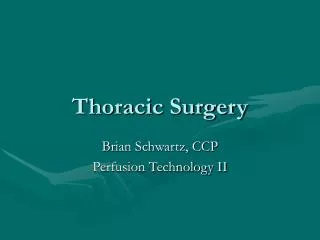
What is the diagnosis? What are the complications of this condition? Is treatment necessary? What treatment is possible ? A barium swallow was performed on an ELDERLY patient who had difficulty in swallowing
WHAT IS IT? Pharyngo-esophageal diverticulum False “pulsion” diverticulum containing mucosa and submucosa Occurs in the neck just above the UES at the pharyngoesophageal junction through Killian’s triangle Dysphagia – Zenker’s Diverticulum
Develops on posterior wall of pharynx between upper and lower divisions of inferior constrictor muscle UES
In most cases the initiating cause is unknown In some cases the cause is GERD related UESspasm ACQUIRED – 80% occur in age >50 yrs Most common esophageal diverticulum WHAT ARE THE SYMPTOMS AND SIGNS? Intermittent cervical dysphagia Gurgling noises in the neck on drinking liquids Food regurgitation Foul breath Left neck swelling Spells of choking Zenker’s Diverticulum
HOW IS THE DIAGNOSIS MADE? Barium Swallow IS IT SERIOUS? Life-threatening due to acute aspiration pneumonia, lung abscess and empyema Disability due to recurrent aspiration pneumonia, fibrosis, bronchiectasis Total dysphagia can occur with large diverticulum distending with retained food causing extrinsic compression Zenker’s Diverticulum
NEED AN OPERATION 1. Cricopharyngeal myotomy in all the cases 2. Management of diverticulum depends on size Small <3 cm: no need for excision Large > 3 cm: add diverticulectomy Concomitant Symptomatic GERD Should be managed first - otherwise risk free aspiration after operation for diverticulum Reason: Reflux is due to incompetent LES. Operation for Zenker’s diverticulum will make UES hypotensive What is the treatment?
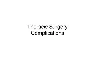
1. What is demonstrated in this barium swallow? 2. What are the essential clinical features? 3. What is necessary to confirm diagnosis? 4. What treatment would you suggest? A 35-year-old man with slowly worsening difficulty swallowing had barium study performed
WHAT IS IT? Esophageal motility disorder characterized by 1.Absence of peristalsis in the body of the esophagus 2.Failure of or incomplete relaxation of LES is response to swallowing, 3. Higher than normal resting LES pressure Dysphagia - Achalasia
WHAT IS THE CAUSE? NA – cause is unknown, viral infection, autoimmune SA – Chagas’ disease due to parasite Trypanosoma Cruzi Finding: degeneration of ganglion cells in Auerbach’s plexus WHAT ARE THE SYMPTOMS? Dysphagia for both solids and liquids; worse with liquids Retrosternal burning discomfort due to food stasis and retention esophagitis Nocturnal regurgitation of food and choking episodes aspiration Achalasia
Esophageal Manometry confirms the diagnosis LES does not relax during swallow Absence of peristalsis
Diagnosis of Achalasia • Suspect diagnosis: from symptoms • Support diagnosis: from esophagogram see “Birds Beak” deformity • Confirm diagnosis: from UGI Endoscopy and Manometry
Achalasia and Epiphrenic Diverticulum Always suspect underlying cause for epiphrenic diverticulum The cause must be treated as well
ESOPHAGUS Malnutrition Progressive dilatation Retention esophagitis Epiphrenic diverticulum Esophageal cancer: squamous (due to retention esophagitis) adenocarcinoma ( due to post treatment reflux – Barrett’s epithelium) RESPIRATORY Aspiration pneumonia, empyema, lung abscess, fibrosis, bronchiectasis Dyspnea due to extrinsic tracheal compression PSYCHOSOCIAL Unable to eat in public withdrawn Complications of Achalasia
What is the treatment for Achalasia? • Chronic condition, no cure for it • Aim of Treatment: relieve distal esophageal functional obstruction • Choices of treatment: • Pneumatic “Balloon” dilatation, initial success rate of 80% decreases to 50% at 10 years; esophageal perforation risk of 5% • Intra-sphincteric injection of Botox, symptomatic relief of 60% decreases to 30% at 2.5 years • Distal esophagomyotomy and partial fundoplication gives the best sustained result of 90%, postoperative reflux is about 15% over time
Distal Esophageal Spasm • “The lower part of the esophagus (smooth muscle) of patients with diffuse esophageal spasm is simultaneously and firmly contracted for an abnormally long time” • Severe pain, dysphagia, and presence of esophageal diverticulae Treatment • Reassurance in most cases • Surgical treatment cannot correct the functional disorder • Long Esophagomyotomy to lower amplitude of waves and resting pressure; add Partial Fundoplication
Nutcracker esophagus • High Amplitude, Peristaltic Esophageal Contractions • > 180 mmHg amplitude • Long duration contractions > 6 sec • LES is normal Treatment • Reassurance in most cases • Must exclude myocardial ischemia • Long Esophagomyotomy in selected cases; add Partial Fundoplication
WHAT IS IT? Frequent retrograde flow of gastric contents across the GE junction into the esophagus WHAT IS THE REASON? Loss of barrier function of the LES, either continuous or intermittent WHAT ARE THE TWO TYPES OF REFLUX? Physiological Pathological – GERD REFLUXATE Acid or Alkaline reflux HCL, Pepsin, Bile, Bile salts Gastroesophageal Reflux Disorder
What are the properties of LES? • Major barrier to reflux – HIGH PRESSURE ZONE • Physiological sphincter • Located in the last 2 to 4 cm of esophagus • Normal resting tone 15 to 30 mm. Hg • Relaxation is coordinated with primary peristalsis • LES pressure is decreased by estrogen, progesterone, nitroglycerine, calcium channel blocker, cigarette smoking, alcohol, fat rich meals, gastric distension, coffee, chocolates, vagotomy, distal esophagomyotomy
WHAT ARE THE CAUSES OF PATHOLOGIC GERD? Idiopathic - majority After pneumatic dilatation or esophagomyotomy for Achalasia Scleroderma Fixed large hiatus hernia Gastric outlet obstruction Prolonged nasogastric tube insertion WHAT ARE THE TYPICAL SYMPTOMS? Unpleasant and intense substernal burning sensation Substernal chest pain Postural and/or postprandial regurgitation Water brash Flatulence Intermittent difficulty with swallowing Lower esophageal sphincter has become incompetent in GERD
ESOPHAGUS - reflux esophagitis: inflammation, erosion and ulceration chronic blood loss and iron deficiency anemia, fibrosis and peptic stricture, Barrett’s epithelium dysplasia adenocarcinoma UES SPASM Zenker’s diverticulum MOUTH - teeth decay and loss of enamel PROXIMAL AIRWAY -laryngitis, wheezing, cough LUNGS - aspiration pneumonia lung abscess, pulmonary fibrosis, bronchiectasis, empyema Complications of Pathologic Gastroesophageal Reflux Disorder
How is the diagnosis of GERD made? • Barium swallow and UGI series • radiologic reflux, hiatus hernia, esophageal stricture, aspiration, spasm in UES • Upper GI endoscopy • esophagitis (erythema, erosions, ulcerations, stricture formation), columnar-lined esophagus • Esophageal manometry • decreased LES, ineffective esophageal peristalsis • 24-hour esophageal pH monitoring • Most sensitive test for acid reflux: number of reflux episodes, duration of reflux, upright vs. supine
FIRST MEDICAL THERAPY Dietary modification Small meals, avoid eating for 2 hrs before going to bed Elevate head of the bed Abstain from coffee, alcohol, trigger foods Drugs: Antacids, PPI, H2- blockers SURGICAL THERAPY IS BY FUNDOPLICATION When GERD is refractory to optimal medical therapy given for a minimum of 6 months When GERD is associated with complications of hiatus hernia, complications in the airway What is the treatment for GERD?
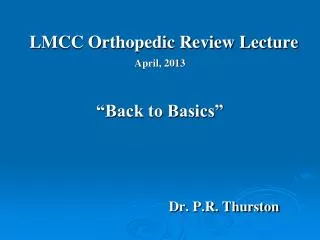
What condition is shown? How does it affect the patient? What serious problem can occur? An elderly patient in the ER complaining of central chest pain radiating into left shoulder, retching, and coffee ground emesis. Barium study from 12 months ago for similar complaint is shown
Complications of Hiatus Hernia 1. Incarceration strangulation ischemic perforation death 2. Anemia – chronic blood loss due to mucosal congestion 3. Dyspnea – large hernia 4. Cardiac Arrhythmias – extrinsic pressure 5. Volvulus obstruction 6. Perforation 7. Massive Bleeding
Type I Type II Type III
Type IV hiatus hernia Intrathoracic stomach with risk of volvulus, associated herniation of transverse colon, small bowel
What are the clinical features of this condition? What is the differential diagnosis? What investigations should be undertaken? What treatments are available? A barium study is finally given to a patient whose complaint for difficulty swallowing was for ignored for 5 months
WHAT ARE THE TWO MAIN CELL TYPES? Adenocarcinoma Squamous cell carcinoma WHAT IS THE MOST COMMON HISTOLOGY? Worldwide: squamous cell carcinoma 95% Western world: adenocarcinoma Esophageal Cancer
Squamous Cell Cancer – what are the etiological factors? • Strong association with excess cigarette smoking and alcohol consumption • Three dietary factors are high intake of nitrosamines (food preservatives), low intake of both vitamin A and nicotinic acid, and chronic iron deficiency • Long standing achalasia, accidental caustic ingestion • Tylosis palmaris et plantaris • Celiac disease • Silica in wheat • Previous radiation therapy to the mediastinum
Adenocarcinoma – what is the cause? • Incidence of adenocarcinoma is rising in NA – an explosion • Due to Chronic GERD – not necessarily acid reflux • Refluxate: acid, pepsin, bile salts, bile • Develops in acquired metaplastic Barrett’s epithelium
Chronic GERD: Adenocarcinoma Mucosal Squamous Epithelium Metaplasia Mucosal Columnar Epithelium “Barrett’s Esophagus” specialized intestinal Dysplasia Low grade High Grade Adenocarcinoma In situ Invasive
WHAT ARE THE SYMPTOMS? Progressive dysphagia, initially for solids and later for liquids Progressive weight loss Means only one diagnosis – Cancer Other symptoms – chest pain, back pain, hoarseness, choking and aspiration, symptoms of metastasis HOW IS THE DIAGNOSIS MADE? Barium UGI series Esophagoscopy and mucosal biopsies HOW IS STAGING DONE? Staging requires CT scan chest and abdomen, Esophageal U/S, PET scan Esophageal Cancer
Treatment for Esophageal Cancer • Surgical: esophagectomy and reconstruction with stomach or colon interposition • Induction chemotherapy and radiotherapy before surgery • Definitive Radiotherapy only: local treatment • Intent: palliation – symptom control • Intent: cure – disease control • Definitive chemotherapy combined with radiation • Esophageal stent: palliation
Benign Tumors of Esophagus • Leiomyoma is the most common • 90% occur in the lower 2/3rd of the esophagus • Grow slowly and cause dysphagia when size becomes >5 cm • Treatment is surgical by enucleation
WHAT IS IT? Bleeding into the pericardial sac, resulting in constriction of right side of the heart, impaired venous return to the heart resulting in decreased stroke volume and cardiac output In acute situation – the pericardium does not stretch and rapid intrapericardial accumulation of even 150mls blood can lead to cardiac tamponade WHAT ARE THE SYMPTOMS AND SIGNS? Suggestive wound Pulse↑, BP↓, JVP↑ Pulsus paradoxus, Kussmaul’s sign DEFINE BECK’S TRIAD Hypotension, Raised JVP, Muffled heart sounds WHAT IS KUSSMAUL’S SIGN? Jugular venous distension with inspiration Immediately life-threatening chest injury - Cardiac Tamponade
HOW IS CARDIAC TAMPONADE DEFINITELY DIAGNOSED IN CHEST TRAUMA? By Echocardiogram Nature of the chest injury Not from CXR – don’t expect to see cardiomegaly WHAT IS THE TREATMENT? Resuscitation protocol for Airway – Breathing –Circulation Immediate IV fluid bolus After pericardiocentesis follow with mandatory surgical exploration Pericardiocentesis is a temporizing procedure Cardiac Tamponade in Chest Trauma
Chest Trauma: Cardiac Tamponade Intrapericardial Pressure (mm Hg)
WHAT IS IT? Bleeding into the pleural space HOW IS IT DIAGNOSED? Hypotension Decreased or absent breath sounds on one or both sides Dullness to percussion CXR, CT scan Chest tube output Immediately life-threatening chest injury - Massive Hemothorax