Download

1 / 40
490 likes | 1.64k Views
Cytology after radical trachelectomy for cervical cancer. Dr Naveena Singh, Edit Titmuss, Dr JoAnne Chin Aleong, Geoffrey Curran, Dr Michael Sheaff, Mr Ian Jacobs, and Mr John Shepherd. Cervical Cancer. Cervical cancer -incidence (per 100,000 women).

E N D
Cytology after radical trachelectomy for cervical cancer Dr Naveena Singh, Edit Titmuss, Dr JoAnne Chin Aleong, Geoffrey Curran, Dr Michael Sheaff, Mr Ian Jacobs, and Mr John Shepherd
Cervical cancer -incidence(per 100,000 women) Where reliable data is available the world wide incidence of cervical cancer varies across continents and from country to country. In the United Kingdom the incidence is about 10 per 100,000. World wide – approx 470, 000 cases & 230,000 deaths
CIN3 - incidence(per 100,000 women) In the UK the incidence of high-grade CIN peaks in the age range 25-29.
Cervical cancer - incidence(per 100,000 women) Whereas the incidence of cervical cancer has a peak at age range at 35-39 followed by a second peak at 75-79 years - probably representing unscreened or poorly screened women.
Cervical cancer -incidence • In 2000 2424 registered cases in UK • About 10 per 100,000 women • In 2002 927 deaths/year in UK • 12th commonest cancer women in UK • 2% of all female cancers • 2nd COMMONEST MALIGNANCY IN WOMEN UNDER 35
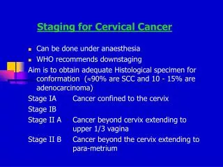
Cervical cancer – stage 1 Outside pelvis Stage I (40%) - confined to cervix
Cervical cancer – stage 2 Stage II (30%) - beyond cervix, not to pelvic side wall or lower 1/3 vagina
Cervical cancer – stage 3 Stage III (25%) - to side wall, lower 1/3 vagina or hydronephrosis
Cervical cancer – stage 4 Stage IV (5%) - involving bladder/rectal mucosa
Cervical cancer - treatment modalities • Surgery • LLETZ/Cone biopsy • Radical Hysterectomy • Exenteration • Radiotherapy • Chemoradiation
Cervical cancer - treatment • Radical hysterectomy, radiotherapy and chemoradiation are all radical modalities • Majority of cancers detected in younger women are early stage • ? too radical for early disease • ? can fertility be conserved
Radical trachelectomy • Dargent et al, 1994 • Cx + parametrium + upper vagina removed • Pelvic lymphadenectomy • Isthmic-vaginal anastomosis • Isthmic cerclage
Radical trachelectomy • Indications • Women under 40 • Cancers up to Stage Ib (IIa) • Strong desire to maintain fertility • Over 90 carried out at St Bartholomew’s Hospital • 3 recurrences and 1 death • 26 live births
Radical trachelectomy -follow-up • CYTOLOGY IS CRUCIAL IN FOLLOW-UP • Isthmic-vaginal smears are taken using brush and spatula • 3 monthly in first year • 4 monthly in second year • 6 monthly from 2-5 years • annually thereafter till 10 years • After 10 years, discharged and sent to NHSCSP call-recall programme
Follow-up • Follow-up for 10-99 months • No of smears/case 1-12 (mean ~ 4) • Interval from surgery to follow-up 2-99 months
Results • 197 smears from 32 patients • Aged 22-36 • Diagnosis - all stage 1B1 • Squamous cell carcinoma 23 • Adenocarcinoma 8 • Adenosquamous 1
Squamous cells • Sole component in 81 smears (41%) from 18 patients, 3-60 months post-operative • 3 cases (10%) contained only squamous cells in all smears • 35 smears (18%) were cytolytic & appeared vaginal • Reason believed to be • High metaplasia • Difficulty in obtaining smear &/or tight os
Keratinisation • Small keratinised cells seen in 29 (15%) of smears • No accompanying features of viral infection • Prominent keratinisation • Large numbers parakeratotic cells • Anucleate squamous cells
Endocervical cells • Typical endocervical cells seen in 48 (28%) of smears • Histology showed ½ cases with endocervical mucosa at upper end of trachelectomy specimen • Generally well preserved • Occasional reactive changes • Multinucleation
Endometrial cells Four types • Functional endometrium • Menstrual endometrium • Lower uterine segment • Tuboendometrial metaplasia
Cytology – Endometrial cells • Present in 114 (58%) of cases. • Small cells with hyperchromatic nuclei • Potential to overcall if not attentive to • Low power arrangement • Two cell population • LMP date • Nature of previous malignancy
Lower uterine segment sampling • LUS sampling feature of brush samples or due to shortening of canal after treatment • Avoid misinterpretation as endometriosis or glandular malignancy • Two cell types • Cuboidal epithelial cells • Tangles of stromal cells • Epithelial microbiospies • Tubular fragments endometrial glands • Branching sheets • 3D clusters • Mitotic figures seen up to day 19
Tubo endometrial metaplasia • Begin histological change in the cervices women treated by cone biopsy or LLETZ for CIN or CGIN • Avoid misinterpreting TEM for CIN or CGIN • Response to injury • Cells smaller than endocervical cells or squamous metaplasia • Centrally or basally situated small round or oval nuclei • Uniformly hyperchromatic with inconspicuous nucleoli • Pseudo-stratification and nuclear crowding or super-crowding • Finely granular evenly dispersed chromatin • Inconspicuous nuclear membrane • Some cells devoid cytoplasm, others with small rim • 2D or 3D with 3D glandular structures with reversed rosettes (nuclei arranged around a clear central zone) • May see terminal bars/cilia • Mitotic figures rare cases • Apoptotic figures and feathering not seen
Stromal fragments • 32/73 (44%) within 12 months after surgery • 33/127 (27%) post-12 months • Heterogeneous in nature • Two types • Plumper type of endometrial origin • Elongated thin wavy nuclei in fragments including capillary vascular channels • Not always possible to categorise
Abnormal Cells As the smears are from a specialist patient population a high referral rate from screeners is to be expected and any smear showing an abnormality should be referred for consultant level opinion
Abnormalities • One fifth slides referred for consultant level opinion • 3 unsatisfactory • 10 borderline • 1 severe dyskaryosis • 3 glandular abnormality • 3 recurrent carcinoma
Recurrences • 3 patients developed recurrent disease • 1 recurrent VAIN 1-2 with koilocytosis • 2 with pelvic recurrences of squamous carcinoma • Both detected in asymptomatic patients
Adequacy • 81 (41%) smears only squamous cells • Do these represent inadequate sampling? • High squamous metaplasia • Difficulty in obtaining smear, tight os • 10% cases never had glandular cells in any smear • Recurrent case detected on proper isthmic smear
Recommendations on adequacy • Smears up to 24 months after trachelectomy must contain glandular cells • If repeat smear is known to have been taken by proper method and is still devoid of glandular cells, report as negative but record absence of glandular cells
False positives 4 (2%) overcalled from 3 patients • 3 severe dyskaryosis • 1 glandular neoplasia • Two reclassified as negative at MDT review • 3rd patient underwent trucut biopsy before slide review
Reporting recommendations • Smears contain only squamous cells • report as unsatisfactory if within 2 years of follow-up and negative thereafter • Abnormal smears • should be followed by smear review and MDT discussion to avoid unnecessary investigations
Conclusion • Recurrence can be detected cytologically before clinical/radiological presentation • Unique in containing large numbers of endometrial cells • Potential for over-diagnosis
Summary • Trachelectomy represents conservative surgical approach for early stage invasive cervical cancer • Likely to increase in popularity • Cytology is mainstay of follow-up • Essentially cytological features are predictable and similar to those after cone biopsy