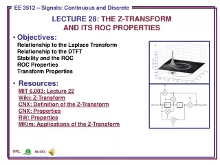
Download
1 / 14
190 likes | 1.34k Views
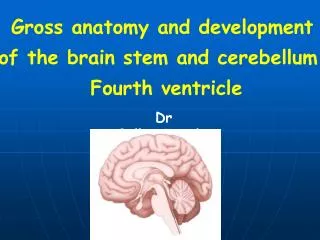
LECTURE 28- ANATOMY OF CEREBELLUM AND ITS CONNECTIONS. Dr. Mohammad Rehan Asad. At the end of the lecture the student should be able to. Identify external features of cerebellum Enumerate neurons, fibers, nuclei and layers of cerebellum Identify connections of cerebellum

E N D
LECTURE 28- ANATOMY OF CEREBELLUM AND ITS CONNECTIONS Dr. Mohammad Rehan Asad
At the end of the lecture the student should be able to • Identify external features of cerebellum • Enumerate neurons, fibers, nuclei and layers of cerebellum • Identify connections of cerebellum • Identify clinical application
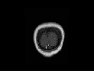
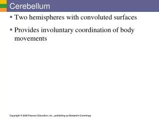
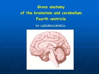
Cerebellum Situated in the posterior cranial fossa and is covered superiorly by the tentorium cerebelli. It is the largest part of the hindbrain (10% of total weight) and lies posterior to the fourth ventricle, the pons, and the medulla oblongata. It consists of two cerebellar hemispheres joined by a narrow median vermis.
Cerebellum Three symmetrical bundles of nerve fibers called the superior, middle, and inferior cerebellar peduncles. Superior peduncle enters mid brain Middle peduncule consist of transverse fibres of pons Inferior peduncle connect with medulla
Cerebellum The cerebellum is divided into three main lobes: the anterior lobe, the middle lobe, and the flocculonodular lobe. The anterior lobe may be seen on the superior surface. It is separated from the middle lobe by a wide V-shaped fissure called the primary fissure. The middle lobe (sometimes called the posterior lobe), which is the largest part of the cerebellum, is situated between the primary and uvulonodular fissures. The flocculonodular lobe is situated posterior to the uvulonodular fissure.
Cerebellum • Sup part of vermis • Lingula • Culmen • Declive • Folium • Inferior part of vermis • Tuber • Pyramid • Uvula • Nodule
Cerebellum connections • Functionally cerebellum divided in corpus cerebelli and flocculonodular lobe • Corpus cerebelli: • afferent from spinal cord and trigeminal nuclei • Inputs from pontine nucleus • Flocculonodular lobe • Connections with vestibular nucleus
Cerebellum connections • Anterior lobe and pyramid mainly receive spinal and trigeminal afferents • Corticopontine connections are relayed to posterior lobe, tuber, vermis and uvula
Cerebellar nuclei Histologically made up of three layers Embedded in white matter are four paired nuclei Dentate is largest Main connection is cerebropontocerebellar Efferent fibres pass to contralateral red nucleus, thalamus, and cerebral cortex.
Cerebellar peduncles • Superior cerebellar peduncle • Efferent of dentate nucleus • Afferent: anterior spinocerebellar tract, Tectocerebellar from mid brain. • Middle cerebellar peduncle • Afferent fibres from pontine nucleus. • Inferior cerebellar peduncle • Efferent: cerebellovestibulartarct • Afferent: vestibulocerebellar tract, post spinocerebellar, cuneocerebellar, oivocerebellar
Blood supply • Posterior inferior cerebellar artery • Anterior inferior cerebellar artery • Branch of basilar artery • Superior cerebellar artery
Clinical application • Lesion of floccolonodular lobe leads to disequilibrium. • Lesion of cerebropontine connections leads to hypotonia, diminished muscle jerk, intention tremor, clumsy movements • Isolated lesions of the vermis are produced in children by medulloblastomas in the roof of the fourth ventricle
Clinical application • Anterior lobe lesions leads to ataxia • Dysdiadochokinesia: inability to perform alternating movements regularly and rapidly • Dysarthria occurs in cerebellar disease because of ataxia of the muscles of the larynx