Quattro Presentation
410 likes | 555 Views
Quattro Presentation. Part I---About Photoluminescence. About Fluorescence. The phenomenon of temporary light absorption and subsequent light emission is called Photoluminescence. The Jablonski Diagram. Absorption (10 -15 s) Internal conversion (10 -12 s)

Quattro Presentation
E N D
Presentation Transcript
About Fluorescence The phenomenon of temporary light absorption and subsequent light emission is called Photoluminescence
The Jablonski Diagram • Absorption (10-15 s) • Internal conversion (10-12 s) • Intersystem crossing (heavy-atom enhanced) • Quenching by external quenchers (diffusion-controlled) • Fluorescence (10-9 s) • Phosphorescence/Inorganic luminescence (10-5 - 101 s) Kasha’s Rule: Photoluminescence always from S1 (fluorescence) or T1 (phosphorescence)
Important Characteristics of Photoluminescence • Each Photoluminescence substance has its own excitation and emission spectra. • The intensity of the Photoluminescence is proportional to its absorption, which in turn is proportional to the excitation light. • The Photoluminescence emission of a substance is always at a higher wavelength than the light used to excite the substance (Stokes-shift phenomenon). • The Photoluminescence of a substance is sensitive to many factors and will change or even disappear under different conditions.
Advantage of Using Photoluminescence as Research Tool • It is very sensitive • It allows to measure very small amount of substance in a given sample • It allows to measure very small changes • It allows to track conformational changes • It is very safe • It is very fast • It is cost-effective • It is widely used in every research
What Can be Measured of a Photoluminescence Substance • Photoluminescence intensity • Excitation spectra • Emission spectra • Photoluminescence lifetime • Anisotropy & Polarization • Time-based Photoluminescence intensity • Time-based Photoluminescence lifetime Quattro II can measure them all!
Part II---General Photoluminescence Instruments and Instruments from PTI/ OBB Family
Photoluminescence System Composition--General • Excitation • Excitation light source • Excitation wavelength selection • Optics for sample excitation • Sample holder/ compartment • Emission • Emission wavelength selection • Optics for sample emission • Detector • Computer interface • Software • Accessories & Attachments
Photoluminescence Systems from OBB/ PTI • Complete Integrated PLSystems: • Quattro • Easylife Series • FluoDia T70 High Temperature fluorescence Plate Reader • Complete open architecture design PL Systems: • QuantaMaster (QM) system for steady-state luminescence measurement • RatioMaster (RM) system for Ratiometric fluorescent measurement of microscopic samples using PMT detector(s) • Easy-Ratio Pro (ERP) system for Ratio fluorescent imaging of microscopic samples using camera as detector • TimeMaster (TM) system for Time-resolved luminescence measurement • Optical Building Blocks: light sources, monochromator, detectors, accessories, et al
Who are We? • PTI is the oldest independent photoluminescence company, specializing in manufacturing and selling complete spectroscopy systems as well as their basic building blocks globally for nearly thirty years. • The Company was established in 1983 in New Jersey, U.S.A. , where the current Headquarter is located • It has a Canadian subsidiary, where most of manufacturing is done and also offices in UK and Germany. • It has international representatives in most countries worldwide. • PTI is entirely dedicated to Photoluminescence products. • In 2007, Optical Building Blocks was established from within PTI -all Integrated Photoluminescence systems, along with optical building blocks now form the core products of Optical Building Blocks Corp., with headquarter in New Jersey, USA
Quattro • Ultra-high sensitivity: S/N > 20,000:1 using water Raman • Single-shot operation • Exceptional precision, stability and reproducibility • Very low stray-light • Super fast acquisitions: measure up to 500 complete decays in a second • Fully automated: complete spectral and lifetime characterization with one click • Easy to operate software. • Low Maintenance • Anti-photo-bleaching design • Ultra-high Sensitivity • Exceptional precision • Super fast speed • Very low stray-light • Extremely easy to use
Unmatchable Sensitivity S/N of 40000:1 measured with Water Raman here on this unit
Super Fast Acquisition Decay of Terbium: t1=422.4us (100%) Offset=-0.000014V, Chi-sq=1.13 (Auto mode, Integration: 10000 Shots at 250 HZ Obtain Decay Data within several seconds.
Optical Layout Light Source Emission Monochromator Detector Excitation Monochromator Sample Compartment
Excitation Light Source • High power pulsed xenon lamp with pre-aligned ellipsoidal reflector • Wavelength Range: 200 to 2000 nm • Pulsed Repetition rate: 1 to 500 HZ (software control ) • Pulse Width 1 µs (FWHM) • Energy per flash: up to 0.15 J • Each pulse is excitation corrected by a silicon photodiode • Lamp life time: 1 x 10⁹ flashes - flashing only during acquisition extends the lamp life time up to 10 years with 8 hr operation per day.
Excitation Monochromator • Asymmetrical Czerny-Turner, coma corrected for minimal stray light • Focal length: 250 mm • Anti-backlashed sine-drive • Precision, two stage auto-calibration • Range: -50 to 1100 nm • Computer selectable spectral bandwidth: 1.5, 2.5, 5, 10, 20 nm slits • Throughput: 70% at 300 nm – grating blazed @ 300nm • Grating type: ruled • Grating size: 50 x 50 mm • Grating groove density: 1200 lines/mm • Apertures: input - f/3.3; output - f/3.7 • Reciprocal linear dispersion: 3.3 nm/mm • Stray light: <0.005% 20 nm away from 632.8 nm Helium Neon line • Accuracy: ±0.2 nm • Optical resolution: 1.5 nm • Maximum scanning speed: 50 nm/sec
Excitation Correction • RCQC: • Type: Enhanced silicon photodiode • Gain: 1 to 1250 times • Linearity: Linear for the entire range of gain values • Range: 200 – 1100 nm • Calibration at factory: 250 – 600 nm vs. rhodamine White: a lamp scan without excitation correction; Red: Same scan with excitation correction activated. Conclusion: standard deviation is better than 0.6 % (260 – 600 nm)
Sample Compartment • Standard Peltier thermo-electrical temperature controlled 3-window cuvette holder: 10° -70° C • Standard Variable speed magnetic stirrer • Aberration corrected demagnified optics result in a light spot <500 um at the sample. • Sample light collection solid angle: 40 • Beam splitter reflects a fraction of the excitation beam to a RCQC (Reference Channel Quantum Counter) photodiode to monitor fluctuations in the excitation beam, both temporal and wavelength. • Double sets of excitation and emission filter holders accommodate 1 inch. diameter round filters • Gas purgeable compartment
Temperature Controlled Sample Holder • Temperature ranges: 10 – 70 °C (nitrogen purging is required when <15°C) • Maximum temperature ramping speed: 20 °C per min (0.33°C/ sec) • Accuracy: ±0.1 °C • Temperature settings resolution: 10 bit (0.06°C) • Temperature stability (after settling): 0.1°C • Water cooled heat sink.
Emission Monochromator • Asymmetrical Czerny-Turner, coma corrected for maximal throughput • Focal length: 250 mm • Anti-backlashed sine-drive • Precision, two stage auto-calibration • Range: -50 to 1100 nm • Computer selectable spectral bandwidth: 1.5, 2.5, 5, 10, 20 nm slits • Throughput: 70% at 500 nm – grating blazed @ 500nm • Grating type: ruled • Grating size: 50 x 50 mm • Grating groove density: 1200 lines/mm • Apertures: input - f/3.7; output - f/3.7 • Reciprocal linear dispersion: 3.3 nm/mm • Stray light: <0.015% 20 nm away from 632.8 nm Helium Neon line • Accuracy: ±0.2 nm • Optical resolution: 1.5 nm • Maximum scanning speed: 50 nm/sec
Detector • Detector: DC Electrical Coupling, • Typical wavelength range 185-650nm • Optional: 185-900 nm • Instrument Response: 6 μs
Instrument Response Function Digitizing: 0.8 us/ point (Fastest) Xe Lamp flash (FWHM): 1.1 us PMT and electronic amplifier response (FWHM): 6 usec Minimum lifetime: 50 usec (Auto Mode). 10 usec. (Manual mode)
Emission & Excitation Correction White: synchronous scan of a lamp without excitation and emission correction ; Green: Same scan with excitation and emission correction activated. Conclusion: standard deviation of synchronous scan is better than 8 % (over Teflon diffuser)
Software and Electronics • Real time digital signal filtering (user selectable) • Transient digitizer acquisition sampling rate: 1.25 Ms/s (0.8 ms/data point) • Digitizing: 16 bits • FluoreScan software • All acquisitions are selected from the main screen of the software. • One click “water Raman” test to confirm system sensitivity • Automatic identification and optimization of excitation and emission wavelengths for unknown samples. • Multiple lifetime fitting 1 to 4 exponentials. • Life times decays can be plotted in real time with automatic exponential fitting
Analysis • Automatic Analysis features can be applied in real time: • Digital filtering, • Automatic multi-exponential fitting. • Math and curve process functions: • Normalize, average, Integrate, differentiate • Multi peak finder, smooth • 3D peak finder • Automatic water Raman calculation
Options • Rotatable solid sample holders • Rotatable powder sample holders • Polarizers • Filters (Long pass, bandpass, neutral Density filters) • Semi-micro cuvette
Data Acquisition • Phosphorescence decay with instant lifetime calculation (up to 4) • Time-based lifetime scan • Anisotropy decay (rotational correlation times) • Lifetime temperature ramping • 3-D excitation vs. decay vs. intensity • 3-D emission vs decay vs intensity • Corrected excitation/ Emission spectra • Time-based intensity scans • Polarization (anisotropy) excitation and emission spectra • Synchronous scans • Intensity vs temperature ramping • 3-D excitation/ emission mapping (EEM): Excitation vs emission vs intensity
Automatic Calibration • FluoreScan has four auto calibration procedures: • Excitation monochromator: for excitation wavelength calibration • Emission monochromator: for emission wavelength calibration • PMT: automatic calculate best HV for the PMT detector for each sample • RCQC: automatic calculate best HV for the detector used for real-time correction for each sample
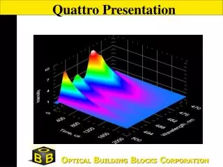
3D Display Real time 3D scan of DYAG : Slits are 2.5 nm; repetition is 250 Hz; HV is 1000V
Applications • Lanthanide-based assays development • LRET based assays • Room temperature phosphorescence of micelle-stabilized aromatics • Room temperature phosphorescence of organics on solid substrate • Optical sensor for carbon dioxide. • Assay for clostridial toxin activity • DNA hybridization assay • Luminescence-based molecular beacons • General steady state fluorescence and phosphorescence • Phosphorescence lifetimes • Inorganic crystals, semiconductors and doped glasses • Protein-protein Interactions • Protein-peptides interactions • Protein folding-unfolding • Membrane Studies • Photosynthesis • Room temperature phosphorescence of proteins (Trp)
Luminescence Excitation and Emission Spectra of Sapphire Crystals
Luminescence Excitation and Emission Spectra of Sapphire Crystals
Luminescence Decay of Sapphire Crystals Deconvolution with Felix 32 Software
Luminescence Decay of Sapphire Crystals Deconvolution with Felix 32 Software
Luminescence Decay of chelated Tb3+ Deconvolution with Felix 32 Software