Molecular Techniques
Molecular Techniques. Studies of cell Fractionation Purification/ Identification Structure/ Function. Proteins. Carbohydrates. Lipids. Nucleic acids. Organelle level. Cell fractionation Nucleus Mitochondria RER, cell membrane SER Cytosol. Cellular level. Microscope.

Molecular Techniques
E N D
Presentation Transcript
Studies of cell • Fractionation • Purification/ Identification • Structure/ Function Proteins Carbohydrates Lipids Nucleic acids Organelle level • Cell fractionation • Nucleus • Mitochondria • RER, cell membrane • SER • Cytosol Cellular level Microscope Molecular level: Macromolecules Atomic level C, H, O, N, S, P
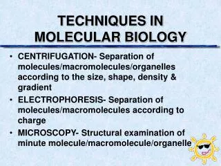
CONTENTS • Cell fractionation • Electrophoresis • Blotting and Hybridization • Polymerase Chain Reaction • DNA Sequences
Cell fractionation • A lab technique which uses a centrifuge to separate the contents of a cell (organelles) into fractions, after the cell has been gently lysed. • The process to break the cells is “HOMOGENIZATION” and the subsequent isolation of organelles is “FRACTIONATION”. • The centrifugation technique is employed to isolate organelles regarding to their physical characteristics, e.g., size, shape and density. The methods frequently used are “DIFFERENTIAL CENTRIFUGATION” and “DENSITY GRADIENT CENTRIFUGRATION”.
HOMOGENIZATION Cell lysis
HOMOGENIZATION • Gently disrupt the cells to release cellular components • Physical or non-physical cell lysis methods • Physical methods of cell disruption • Disruption of cells results from the shearing forces generated between the cells and either solid abrasive or liquid medium. • Pastel and mortar homogenizer, Abrasive beads, Blender, Pressure homogenization, Osmotic shock, Freezing/ thawing technique, Ultrasonification • Non-physical methods of cell disruption • Organic solvents- to destroy membrane • Chaotropic anions- to destabilize membrane • Detergents- to dissolve proteins and lipids membrane • Enzymatic digestion- to digest proteins, carbohydrates and lipids of cell wall
FRACTIONATION Centrifugation
Cell Fractionation • Physical methods of cell disruption • Pastel and mortar homogenizer • Abrasive beads-sands, silica, alumina • Blender-special designed blades and chamber • Pressure homogenization-cells are imbibed with an inert gas (argon) which will form gas bubbles inside cytoplasm when the cells are suddenly returned to atmospheric pressure, hence rupture the membrane. • Osmotic shock-swelling and disrupting of cells in hypotonic solution • Freezing/ thawing technique-ice crystals rupture the cells • Ultrasonification- ultrasonic wave to break open the plasma membrane and leave the internal organelles intact.
Cell Fractionation • Non-physical methods of cell disruption • Organic solvents- chloroform/methanol mixtures can dissolve membrane lipids (destroy membranes) and release subcellular components. • Chaotropic anions- potassium thiocyanate, potassium bromide, lithium diiodosalicylate act to destabilize lipid membranes consequently, the subcellular components are being released. • Detergents- solubilize the integral membrane proteins by interacting with the phospholipid bilayer, e.g., SDS (anionic), Deoxycholate (non-denaturing) and Triton X-100 (non-ionic) • Enzymatic digestion- to digest proteins, carbohydrates and lipids of cell wall, Mixture of enzymes: chitinases, pectinases, lipases, proteases, cellulases
Cell Fractionation • Homogenization medium • Slightly hypo-osmotic or iso-osmotic – to preserve structural integrity of organelles • Osmoticums: sucrose, manitol, sorbitol • Chelating agents: EDTA or EGTA (remove Ca2+ or Mg2+ which are required by membrane proteases) • Protease inhibitor: endopeptidases, exopeptidases • The homogenization should be performed at 4oC to minimize protease activity
Cell Fractionation • Fractionation • The most widely used technique for fractionating cellular components is centrifugation technique • Particles of different density, size, and shape sediment at different rate in a centrifugal field. • Factors affected the rate of sedimentation: • particle size and shape • the viscosity of suspending medium • centrifugal field • * The particle remain stationary when the density of the particle and the density of the centrifugation medium are equal
Cell Fractionation • Types of Centrifugation • Differential centrifugation • Rate-zonal centrifugation • Isopycnic centrifugation A centrifuge working at speeds in excess of 20,000 RPM is an “ultracentrifuge”.
Cell Fractionation • Differential centrifugation • Separates particles as a function of size and density • A particular centrifugal field is chosen over a period of time • Larger mass; lower centrifugation force; lesser spin time • Subjected to repeated steps with increasing of centrifugation force
Differential centrifugation Centrifugation force to pellet the cellular components
Differential Centrifugation Pellet 1 Pellet 2 Pellet 3 Pellet 4
Molecules separate according to size and shape centrifugation Rate-zonal Centrifugation • Rate-zonal centrifugation • Medium • Slightly viscous • Density gradient;a positive increment in density • e.g., sucrose, GuHCl • Sample is applied on the top of density gradient • Particles separate into a series of bands (zone) in accordance to rate of sedimentation (S), size and shape
Centrifugation Fraction collection Rate-zonal Centrifugation
Isopycnic Centrifugation • Isopycnic • centrifugation • Based solely on the density of the particles • Separation medium– self-generating density gradient medium (CsCl medium) • Unaffected by the size or the shape of the particles • Mostly used to separate nucleic acids, large glycoproteins
What make rate-zonal and isopycnic centrifugations difference?
Electrophoresis E v F + - + - - - - + q f • Molecules are separated by electric force • F = qE : where q is net charge, E is electric field strength • The velocity is encountered by friction • qE = fv : where f is frictional force, v is velocity • Therefore, mobility per unit field (U) = v/q = q/f = q/6pr : where is viscosity of supporting medium, r is radius of sphere molecule
Electrophoresis - • Factors affected the mobility of molecules • 1. Molecular factors • Charge • Size • Shape • 2. Environment factors • Electric field strength • Supporting media (pore: sieving effect) • Running buffer +
Electrophoresis • Types of supporting media • Paper • Agarose gel (Agarose gel electrophoresis) • Polyacrylamide gel (PAGE) • pH gradient (Isoelectric focusing electrophoresis) • Cellulose acetate
Agarose Gel • purified large MW polysaccharide (from agar) • very open (large pore) gel • used frequently for large DNA molecules
Electrophoresis Agarose gel staining Ethidium bromide Fluorescence dye Pounseur-S dye
Electrophoresis • Polyacrylamide Gels • Acrylamide polymer; very stable gel • can be made at a wide variety of concentrations • gradient of concentrations: large variety of pore sizes (powerful sieving effect)
Electrophoresis SDS-Polyacrylamide Gel Electrophoresis (SDS-PAGE) • Sodium Dodecyl Sulfate = Sodium Lauryl Sulfate: CH3(CH2)11SO3- Na+ • Amphipathic molecule • Strong detergent to denature proteins • Binding ratio: 1.4 gm SDS/gm protein • Charge and shape normalization
Electrophoresis • Isoelectric Focusing Electrophoresis (IFE) • Separate molecules according to their isoelectric point (pI) • At isoelectric point (pI) molecule has no charge (q=0), hence molecule ceases • pH gradient medium
Electrophoresis • 2-dimensional Gel Electrophoresis • First dimension is IFE (separated by charge) • Second dimension is SDS-PAGE (separated by size) • So called 2D-PAGE • High throughput electrophoresis, high resolution
2-dimensional Gel Electrophoresis • Spot coordination • pH • MW
Hybridization • Can be DNA:DNA, DNA:RNA, or RNA:RNA (RNA is easily degraded) • Dependent on the extent of complementation • Dependent on temperature, salt concentration, and solvents • Small changes in the above factors can be used to discriminate between different sequences (e.g., small mutations can be detected) • Probes can be labeled with radioactivity, fluorescent dyes, enzymes, etc. • Probes can be isolated or synthesized sequences
Oligonucleotide probes • Single stranded DNA (usually 15-40 bp) • Degenerate oligonucleotide probes can be used to identify genes encoding characterized proteins • Use amino acid sequence to predict possible DNA sequences • Hybridize with a combination of probes • TT(T/C) - TGG - ATG - GA(T/C) - TG(T/C) - could be used for FWMDC amino acid sequence • Can specifically detect single nucleotide changes
Detection of Probes • Probes can be labeled with radioactivity, fluorescent dyes, enzymes. • Radioactivity is often detected by X-ray film (autoradiography) • Fluorescent dyes can be detected by fluorometers, scanners • Enzymatic activities are often detected by the production of dyes or light (x-ray film)
RNA Blotting (Northerns) • RNA is separated by size on a denaturing agarose gel and then transferred onto a membrane (blot) • Probe is hybridized to complementary sequences on the blot and excess probe is washed away • Location of probe is determined by detection method (e.g., film, fluorometer)
Applications of RNA Blots Detect the expression level and transcript size of a specific gene in a specific tissue or at a specific time. Sometimes mutations do not affect coding regions but transcriptional regulatory sequences (e.g., UAS/URS, promoter, splice sites, copy number, transcript stability, etc.)
Western Blot • Highly specific qualitative test • Can determine if above or below threshold • Typically used for research • Use denaturing SDS-PAGE • Solubilizes, removes aggregates & adventitious proteins are eliminated Components of the gel are then transferred to a solid support or transfer membrane weight Paper towel Wet filter paper Transfer membrane Paper towel
Add antibody against yours with a marker (becomes the antigen) Stain the bound antibody for colour development It should look like the gel you started with if a positive reaction occurred Western Blot • Block membrane e.g. dried nonfat milk Rinse with ddH2O Add monoclonal antibodies Rinse again Antibodies will bind to specified protein
PCR • A simple rapid, sensitive and versatile in vitro method for selectively amplifying defined sequences/regions of DNA/RNA from an initial complex source of nucleic acid - generates sufficient for subsequent analysis and/or manipulation • Amplification of a small amount of DNA using specific DNA primers (a common method of creating copies of specific fragments of DNA) • DNA fragments are synthesized in vitro by repeated reactions of DNA synthesis (It rapidly amplifies a single DNA molecule into many billions of molecules) • In one application of the technology, small samples of DNA, such as those found in a strand of hair at a crime scene, can produce sufficient copies to carry out forensic tests. • Each cycle the amount of DNA doubles
Background on PCR • Ability to generate identical high copy number DNAs made possible in the 1970s by recombinant DNA technology (i.e., cloning). • Cloning DNA is time consuming and expensive • Probing libraries can be like hunting for a needle in a haystack. • Requires only simple, inexpensive ingredients and a couple hours • PCR, “discovered” in 1983 by Kary Mullis, • Nobel Prize for Chemistry (1993). • It can be performed by hand or in a machine called a thermal cycler.
Three Steps • Separation Double Stranded DNA is denatured by heat into single strands. • Short Primers for DNA replication are added to the mixture. • Priming DNA polymerase catalyzes the production of complementary new strands. • Copying The process is repeated for each new strand created • All three steps are carried out in the same vial but at different temperatures
Step 1: Separation • Combine Target Sequence, DNA primers template, dNTPs, Taq Polymerase • Target Sequence 1. Usually fewer than 3000 bp 2. Identified by a specific pair of DNA primers- usually oligonucleotides that are about 20 nucleotides • Heat to 95°C to separate strands (for 0.5-2 minutes) • Longer times increase denaturation but decrease enzyme and template Magnesium as a Cofactor Stabilizes the reaction between: • oligonucleotides and template DNA • DNA Polymerase and template DNA
Heat Denatures DNA by uncoiling the Double Helix strands.
Step 2: Priming • Decrease temperature by 15-25 ° • Primers anneal to the end of the strand • 0.5-2 minutes • Shorter time increases specificity but decreases yield • Requires knowledge of the base sequences of the 3’ - end
Selecting a Primer • Primer length • Melting Temperature (Tm) • Specificity • Complementary Primer Sequences • G/C content and Polypyrimidine (T, C) or polypurine (A, G) stretches • 3’-end Sequence • Single-stranded DNA
Step 3: Polymerization • Since the Taq polymerase works best at around 75 ° C (the temperature of the hot springs where the bacterium was discovered), the temperature of the vial is raised to 72-75 °C • The DNA polymerase recognizes the primer and makes a complementary copy of the template which is now single stranded. • Approximately 150 nucleotides/sec
Potential Problems with Taq • Lack of proof-reading of newly synthesized DNA. • Potentially can include di-Nucleotriphosphates (dNTPs) that are not complementary to the original strand. • Errors in coding result • Recently discovered thermostable DNA polymerases, Tth and Pfu, are less efficient, yet highly accurate.
How PCR works Begins with DNA containing a sequence to be amplified and a pair of synthetic oligonucleotide primers that flank the sequence. Next, denature the DNA at 94˚C. Rapidly cool the DNA (37-65˚C) and anneal primers to complementary s.s. sequences flanking the target DNA. Extend primers at 70-75˚C using a heat-resistant DNA polymerase (e.g., Taq polymerase derived from Thermus aquaticus). Repeat the cycle of denaturing, annealing, and extension 20-45 times to produce 1 million (220) to 35 trillion copies (245) of the target DNA. Extend the primers at 70-75˚C once more to allow incomplete extension products in the reaction mixture to extend completely. Cool to 4˚C and store or use amplified PCR product for analysis.